Toggle light / dark theme

Smart fluorescent molecules provide cheaper path to sharper microscopy images

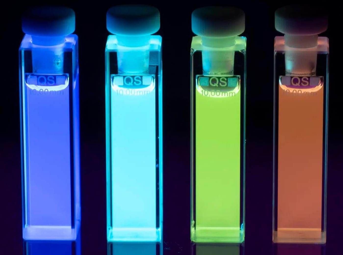
Multiphoton microscopy is used in biomedical research to study cells and tissues. Today, so-called two-photon microscopy is used to study processes within cells, but the technique has limitations in terms of image resolution. Four-photon microscopy provides images with higher resolution. However, such instruments are very expensive and, when studying biological material, the powerful laser light required can damage samples.

“In this project, we have developed molecules to visualize molecular-level details and monitor processes using the more common two-photon microscopy technique. These molecules have the capacity to achieve higher resolution than with four-photon microscopy, although two-photon microscopy is used,” says the project coordinator Joakim Andréasson, Professor at the Department of Chemistry and Chemical Engineering at Chalmers University of Technology.

“In the long term, results from studies of this kind may provide new insights into diseases, pharmaceuticals and the very smallest building blocks of life.”

A rewritable DNA hard drive may help solve the growing data storage crisis

Around the world, scientists are exploring an unexpected solution to the growing data crisis: storing digital information in synthetic DNA. The idea is simple but powerful—DNA is one of the most compact, durable information systems on Earth. But one issue has held the field back. Once data is written into DNA, it can’t be changed.

Now, researchers at the University of Missouri are helping to solve that problem by transforming DNA from a one-time medium into a rewritable digital hard drive. Their research is published in the journal PNAS Nexus.

“DNA is incredible—it stores life’s blueprint in a tiny, stable package,” said Li-Qun “Andrew” Gu, a professor of chemical and biomedical engineering at Mizzou’s College of Engineering. “We wanted to see if we could store and rewrite information at the molecular level faster, simpler and more efficiently than ever before.”

Humanization and engineering of protective antibodies targeting severe fever with thrombocytopenia syndrome virus Gn protein

Ren et al. humanize and structurally optimize a chimeric anti-SFTSV antibody, generating variants with markedly enhanced neutralization potency by strengthening binding to recombinant Gn and intact virions, providing full in vivo protection and establishing a generalizable framework for therapeutic antibody engineering.

Engineered protein markers read living brain gene activity in monkeys via blood

Gene therapy has been successfully used to treat a number of diseases, including immune deficiencies, hereditary blindness, hemophilia and, recently, Huntington’s disease, a fatal neurological disorder.

An advance reported in the journal Neuron adds to the technique’s growing track record of evidence supporting the view that it could unlock powerful, personalized therapies: Rice University bioengineer Jerzy Szablowski and collaborators in Vincent Costa’s lab at Emory University found that released markers of activity (RMAs) — engineered proteins designed to cross the blood-brain barrier and persist in the blood for hours at a time, providing a reliable and noninvasive way to get information about gene expression in the brain — work just as well in monkeys as they do in mice.

On the route from laboratory discovery to lifesaving treatment, large animal model studies are a critical part of the process. Most research never reaches this stage.

Bioengineered neuronal ‘circuit board’ mimics conditions of the human brain

A new bioengineered neuronal circuit board “BioConNet” allows scientists to artificially engineer human brain-like wiring at scale and can be used to engineer any possible circuit. The fully programmable, open-source system allows generation of large-scale circuits, while maintaining the ability to focus on single connections between neurons.

This is a key advance in engineering human-like neural circuits as it allows for a new level of wiring complexity compared to previous systems. BioConNet allows scientists increased control over wiring in the culture compared to existing methods such as organoids and commercially available systems. The research is published in the journal Advanced Healthcare Materials.

“By combining engineering and neurobiology with the most recent stem cell culture techniques, we can now create human-specific, functional, large-scale complex neural circuits in the lab,” said senior author, Dr. Andrea Serio, Reader in Neural Tissue Engineering, Group Leader at the UK Dementia Research Institute (UK DRI) at King’s and Senior Group Leader at the Crick.

Why do microbes team up? A new model explains nutrient sharing in fluctuating environments

Depending on others for something you need may feel like a risky proposition—and perhaps a human one. It is actually a survival strategy found in the microbial world, and far more frequently than one might expect. Discovering why is key to understanding how microbes form stable communities across medical, industrial, and ecological settings.

A new study by bioengineering professor Sergei Maslov (CAIM co-leader), computational scientist Ashish George, and biology professor Tong Wang explores why interdependence can be such a winning move for microbial communities. Their work, published in Cell Systems, demonstrated that a mathematical model of how bacteria produce and share resources accurately predicted the outcome of experiments with living E. coli strains.

The researchers’ collaboration began during their time as colleagues at the Carl R. Woese Institute for Genomic Biology at the University of Illinois Urbana-Champaign. George continued the collaboration in his position at the Broad Institute; Wang, in his appointment at Purdue University. Maslov, who led the study, remains at Illinois and is an affiliate member of the National Institute for Theory and Mathematics in Biology.

Genetic defect that weakens esophageal lining identified!

But the molecular factors responsible for the onset of Barrett’s esophagus remain poorly understood.

The findings, published in Nature Communications, combined family studies, laboratory experiments and genetically engineered mouse models to identify and understand how genetic defects contribute to disease development.

The team sequenced and analyzed genetic material of 684 people from 302 families where multiple members developed Barrett’s esophagus or esophageal cancer. They discovered that a subset of affected family members carry inherited mutations in a gene called VSIG10L.

“We found that this gene acts like a quality control system for the esophageal lining,” said the lead researcher. “When it’s defective, the cells do not mature properly and the protective barrier in the esophageal lining becomes weak, allowing stomach bile acid to cause tissue changes that enhances the risk of developing Barrett’s esophagus.”

When researchers genetically engineered mice with human-equivalent VSIG10L mutations, they found that the esophageal lining became disrupted structurally and molecularly, according to the author. The study found that when the mice were exposed to bile acid, they developed Barrett’s-like disease over time, effectively replicating the disease’s progression in humans.

These genetically engineered mice also represent the first animal model for Barrett’s esophagus based directly on human genetic predisposition to the disease, the author said.

With VSIG10L shown to be a key gene in maintaining esophageal health, family members can now be screened for genetic variants to identify those at a high-risk of developing Barrett’s esophagus or esophageal cancer. ScienceMission sciencenewshighlights.

How chronic inflammation rewires macrophages

TIL therapy for glioblastoma.

Tumor infiltrating lymphocyte (TIL) therapy has demonstrated encouraging efficacy in melanoma and nonsmall-cell lung cancer (NSCLC), and is now being explored for glioblastoma despite its immunologically ‘cold’ microenvironment.

Recent studies confirm that functional TILs can be expanded from cold tumors such as glioblastoma, including solid tumor resections and aspirates, overcoming previous feasibility concerns.

Advances in cytokine support, gene editing, and artificial antigen-presenting cells (APCs) are improving TIL persistence, cytotoxicity, and manufacturing scalability.

Focused ultrasound and nanoparticle delivery offer innovative solutions to enhance TIL infiltration across the blood– brain barrier. Integration of spatial multi-omics enables high-resolution mapping of immune niches and identification of tumorreactive clones.

Combination strategies with checkpoint blockade, myeloid modulation, and oncolytic virotherapy are emerging as rational paths to enhance TIL efficacy sciencenewshighlights ScienceMission https://sciencemission.com/TIL-therapy-17895


Post-Humans of All Tomorrows-3D Size Comparison

In All Tomorrows by C. M. Kosemen, also known as Nemo Ramjet, humanity’s distant descendants are reshaped across millions of years into wildly divergent “post-human” species after being genetically engineered by the godlike alien Qu. These forms range from tiny, almost vermin-like organisms and sessile, colony-bound beings to aquatic leviathans, aerial gliders, and towering, heavily built giants as each adapted to extreme planetary environments and radically different evolutionary pressures. Some retain echoes of recognizable humanity, while others are so transformed they blur the line between animal, ecosystem, and living architecture. In this size comparison, we’ll explore the full spectrum of these post-human forms, from the smallest engineered remnants to the most massive macro-organic descendants.

Credits:
https://all-tomorrows.fandom.com/wiki/Qu.
https://speculativeevolution.fandom.com/wiki/All_Tomorrows.

FAIR-USE COPYRIGHT DISCLAIMER Copyright Disclaimer under Section 107 of the Copyright Act 1976, allowance is made for “fair use” for purposes such as criticism, commenting, news reporting, teaching, scholarship, and research. Fair use is a use permitted by copyright statute that might otherwise be infringing. Non-profit, educational or personal use tips the balance in favour of fair use. Nutbug does not own the rights to these videos and pictures. They have, in accordance with fair use, been repurposed with the intent of educating and inspiring others. However, if any content owners would like their images removed, please contact us by email [email protected]

/* */