Toggle light / dark theme

Robotic microfluidic platform brings AI to lipid nanoparticle design

AI has designed candidate drugs for antibiotic-resistant infections and genetic diseases. But efforts to incorporate AI into the design of lipid nanoparticles (LNPs), the revolutionary delivery vehicles behind mRNA therapies like the COVID-19 vaccines, have been much more limited.

Designing LNPs is especially challenging: Each formulation combines multiple lipid components whose ratios influence how the particle delivers genetic instructions inside cells. Scientists still lack a clear map connecting those chemical inputs to biological outcomes.

The reason? There simply isn’t enough data.

Mapping 3D-super-enhancers with machine learning to pinpoint regulators of cell identity

Scientists usually study the molecular machinery that controls gene expression from the perspective of a linear, two-dimensional genome—even though DNA and its bound proteins function in three dimensions (3D). To better understand how key components of this machinery, such as super-enhancers, regulate genes in this 3D reality, scientists at St. Jude Children’s Research Hospital have developed a new algorithm called BOUQUET.

Using machine learning, BOUQUET reveals that sets of genes and their regulatory elements can interact within protein condensates, high-density membraneless droplets, in cells’ nuclei. The findings, which provide new insight into how cells regulate the genes that control their specialized identities, were published today in Nucleic Acids Research.

Cells express certain sets of genes to carry out specific functions; for example, a blood cell and a brain cell express different context-specific genes. There are 3 billion base pairs of human DNA, and the genes involved in cell identity are scattered throughout. Even more challenging, enhancers, DNA elements that activate gene expression, can be thousands of DNA bases away from their target genes.

Ultrafast light pulses make molecules rotate on quantum materials

Researchers from Germany, Japan and India, led by scientists from DESY and the Universities of Kiel and Hamburg, have found a way to collectively make molecules on a flat surface rotate by exposing them to light using ultrafast light pulses from DESY’s free-electron laser FLASH and a high-harmonic generation source. However, making those molecules dance is not the ultimate goal: this result could have an impact on next-generation quantum and energy materials for electronics, data storage and energy conversion.

Molecules sitting on a material surface usually do just that—they sit on the surface without changing. If you send energy their way, however—for example, in the form of light—they can become dynamic and move. If this movement could be controlled, it could have a massive influence on all sorts of nanomaterials that are being investigated for a variety of applications from health to data storage.

DESY scientist Markus Scholz, leader of a study now published in Nature Communications, points out that this is particularly interesting in hybrid systems where organic molecules are placed on atomically thin, two-dimensional quantum materials. Examples of these hybrid systems are molecular electronics or energy-driven functional surfaces.

Why simulating an entire cell cycle took years, multiple GPUs and six days per run

By simulating the life cycle of a minimal bacterial cell—from DNA replication to protein translation to metabolism and cell division—scientists have opened a new frontier of computer vision into the essential processes of life. The researchers, led by chemistry professor Zan Luthey-Schulten at the University of Illinois Urbana-Champaign, present their findings in the journal Cell.

The team simulated a living cell at nanoscale resolution and recapitulated how every molecule within that cell behaved over the course of a full cell cycle. The work took many years: vast computer resources, large experimental datasets, a suite of experimental and computational techniques and an understanding of the roles, behaviors and physical interactions of thousands of molecular players.

The researchers had to account for every gene, protein, RNA molecule and chemical reaction occurring within the cell to recreate the timing of cellular events. For example, their model had to accurately reflect the processes that allow the cell to double in size prior to cell division.

One-hour saliva test spots biomarker linked to several cancers

QUT researchers have developed a simple one-hour saliva test for a protein biomarker that has been linked with oral, colon and pancreatic cancers. The findings are published in the journal Talanta.

The paper is titled “Label free paper sensor and light driven material for the rapid screening of S100P cancer biomarker in saliva.” Corresponding author, Associate Professor Emad Kiriakous, from QUT’s School of Chemistry and Physics, said this technology could pave the way for simple, low-cost, point-of-care screening tools to help identify and treat cancer early.

Professor Kiriakous said the QUT team developed a rapid testing technique of saliva using paper coated in gold and silver nanoparticles to create a highly sensitive sensor that records the Raman spectrum (or SERS, the process by which a substance scatters laser light which is used to identify molecules) of saliva samples.

This new blood test could detect cancer before it shows up on scans

A new CRISPR-powered light sensor can detect the faintest molecular signs of cancer in a drop of blood. A new light-based sensor can spot incredibly tiny amounts of cancer biomarkers in blood, raising the possibility of earlier and simpler cancer detection. The technology merges DNAnanotechnology, CRISPR, and quantumdots to generate a clear signal from just a few molecules. In lung cancer tests, it worked even in real patient serum samples. Researchers hope it could eventually power portable blood tests for cancer and other diseases.

Scientists have designed a powerful light based sensor capable of detecting extremely small amounts of cancer biomarkers in blood. The innovation could eventually allow doctors to identify early warning signs of cancer and other diseases through a routine blood draw.

Biomarkers such as proteins, fragments of DNA, and other molecules can signal whether cancer is present, how it is progressing, or a person’s risk of developing it. The difficulty is that in the earliest stages of disease, these markers exist in extremely low concentrations, making them hard to measure with conventional tools.

Copper Single-Atoms Loaded on Molybdenum Disulphide Drive Bacterial Cuproptosis-Like Death and Interrupt Drug-Resistance Compensation Pathways

111. Wenqi Wang, Xiaolong Wei, Bolong Xu, Hengshuo Gui, Yan Yan*, Huiyu Liu* & Xianwen Wang* Nano-Micro Lett. 18,111 (2026).

This work is led by Prof. Dr. Xianwen Wang (Anhui Medical University) and co-workers. Prof. Wang’s research centers on burn wounds and tissue regeneration, burn infection, design and development of antimicrobial nanomaterials, development of anti-inflammatory nano-formulations and study on their anti-inflammatory mechanisms. This article develops copper single-atom-loaded MoS₂ nanozymes (Cu SAs/MoS₂) that combat drug-resistant bacteria through a triple mechanism of oxidative damage, cuproptosis-like death, and disrupted cell wall synthesis. Density functional theory reveals that Cu coordination enhances H₂O₂ adsorption, reducing activation energy by 17% and boosting peroxidase-like activity, while glutathione peroxidase-like activity disrupts redox homeostasis and inhibition of peptidoglycan synthesis blocks cell wall remodeling, collectively enabling efficient bacterial killing and decelerating resistance development.

Related articles: Cactus Thorn-Inspired Janus Nanofiber Membranes as a Water Diode for Light-Enhanced Diabetic Wound Healing https://doi.org/10.1007/s40820-025-01904-z Synergistic Ferroptosis–Immunotherapy Nanoplatforms: Multidimensional Engineering for Tumor Microenvironment Remodeling and Therapeutic Optimization https://doi.org/10.1007/s40820-025-01862-6 Wearable Ultrasound Devices for Therapeutic Applications https://doi.org/10.1007/s40820-025-01890-2


The development of highly efficient and multifunctional nanozymes holds promise for addressing the challenges posed by drug-resistant bacteria. Here, copper single-atom-loaded MoS2 nanozymes (Cu SAs/MoS2) were developed to effectively combat drug-resistant bacteria by synergistically integrating the triple strategies of oxidative damage, cuproptosis-like death and disruption of cell wall synthesis. Density functional theory revealed that each Cu center coordinated with three sulfur ligands, enhancing the adsorption of H2O2, which reduced the activation energy of the key step by 17%, thereby improving peroxidase-like (POD-like) activity. The generation of reactive oxygen species in combination with Cu SAs/MoS2 glutathione peroxidase-like (GSH-Px-like) for glutathione scavenging resulted in an imbalance in redox homeostasis within bacteria.

Scientists Spin Molecules Inside a Frictionless Superfluid for the First Time

A newly designed optical centrifuge allows scientists to control molecular rotation inside superfluid helium nano-droplets. Physicists have developed a new version of an optical centrifuge that can control how molecules rotate while they are suspended inside liquid helium nano-droplets. The advan

Ribosome organization during stress!

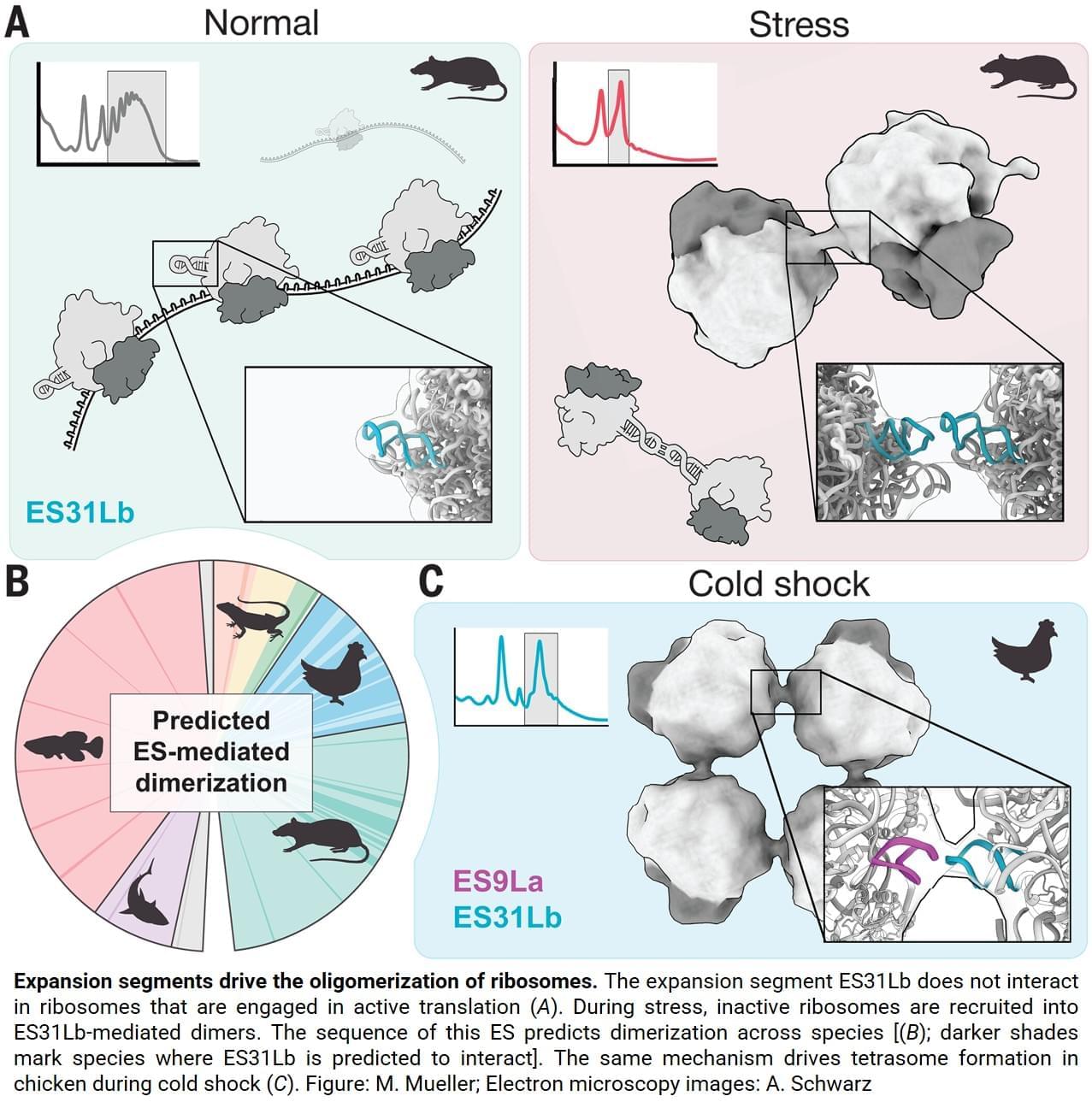
“Surprisingly, the two ribosomes are not held together by proteins, as is common in bacteria. Instead, the connection is made by a specific piece of ribosomal RNA called an expansion segment”, explains one of the lead authors.

Expansion segments are long, flexible RNA “tentacles” that protrude from ribosomes and have grown larger over the course of evolution. Although they are a prominent feature of animal ribosomes, their functions only just started to emerge. This study now shows that one particular expansion segment, called “31b”, is both necessary and sufficient to link ribosomes together during stress. At the molecular level, the expansion segment forms a precise RNA-RNA interaction — a so-called “kissing loop” — in which identical RNA loops bind each other through complementary sequences. Disrupting this interaction prevents disome formation, stunts cellular growth and makes cells more sensitive to stress. Science Mission sciencenewshighlights.


Ribosomes, the cell’s protein-making factories, consume large amounts of energy as they build the proteins that keep cells alive and functioning. When cells experience stress — such as lack of nutrients or sudden drops in temperature — they quickly switch into survival mode. New research now reveals an unexpected way cells manage this transition: By pairing up inactive ribosomes using a ribosomal RNA link. This RNA-based mechanism reveals a previously unknown role for ribosomal RNA in the cellular stress response.

Ribosomes are large molecular machines made of protein and RNA that build all proteins in the cell. Because protein production is extremely energy-intensive, cells rapidly reduce protein synthesis when stressed. It has long been known that bacterial cells pair their inactive ribosomes into so-called “hibernating disomes” however, such structures had not previously been identified in animal cells.

Using advanced imaging techniques, the team discovered that stressed animal cells — including neurons — assemble inactive ribosomes into tightly linked pairs, known as disomes. These ribosome pairs are not accidental collisions or artifacts, but a regulated and reversible response to stress. The new study was published in Science.

Thermogenetics: How proteins are controllable by heat

Protein activity can be precisely regulated via subtle changes in temperature using heat-sensitive switches. Underlying this capability is a novel modular design strategy developed by researchers at the Institute of Pharmacy and Molecular Biotechnology of Heidelberg University. The strategy allows the integration of sensory domains in various proteins regardless of function or spatial structure.

This new approach in the field of thermogenetics is broadly applicable and opens up new possibilities for precise, non-invasive control of different cellular processes. It was developed by a research team led by Prof. Dr. Dominik Niopek and Dr. Jan Mathony and is published in Nature Chemical Biology

Proteins are the molecular machines of the cell. They regulate nearly all vital processes and their responses are highly dynamic. To better understand these processes and their chronological sequence, scientists need tools that can be used to change individual parameters precisely and in a controlled manner. The most suitable proteins are those that can be turned on and off like technical devices. Especially attractive in this context are heat-sensitive protein switches that tightly regulate the temperature spatiotemporally and are able to deeply penetrate tissue or complex biological samples as a signal.

/* */