Toggle light / dark theme

DNA shape explains crucial gene-therapy challenges

CRISPR is a powerful DNA-editing tool that has underpinned huge advancements in human health care in the last decade. It is a precision tool, but is not perfect, and misplaced DNA edits can compromise safety and efficacy, costing billions each year. Researchers at the MRC Laboratory of Medical Sciences (LMS), Imperial College London and the University of Sheffield have published research in Nature showing that the physical twisting of DNA plays an important role in these mistakes. Using a newly developed platform of tiny (nanometer-sized) DNA circles, called DNA minicircles, the team captured never-before-seen interactions between CRISPR and DNA, providing insights that could help eradicate errors altogether.

CRISPR-Cas9 has transformed biology by giving scientists a programmable way to cut and edit DNA. Its ever-growing impact includes groundbreaking therapies for genetic diseases such as sickle cell anemia and an increasing role in personalized cancer treatment and rapid diagnostics. But even carefully designed CRISPR systems can sometimes cut DNA sequences that were not the intended targets.

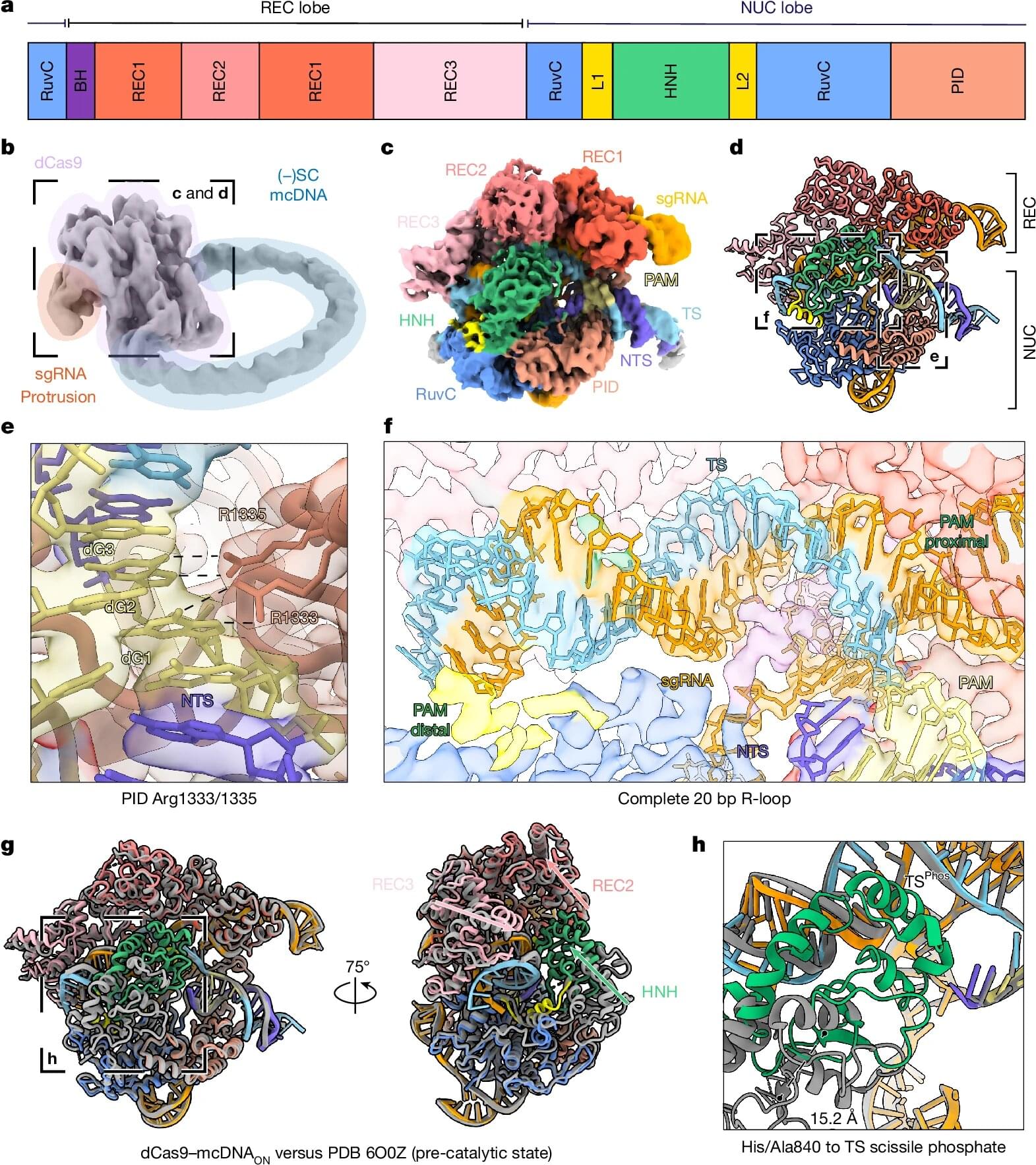
“It’s a tool that is not perfect and can introduce errors and make edits where it shouldn’t make them,” says Professor David Rueda, head of the Single Molecule Imaging group at the LMS and Chair in Molecular and Cellular Biophysics at Imperial College London. “And it’s an important problem for the industry. It’s been estimated to be $0.3 to $0.9 billions per year in industry costs, in profiling off-targets, redesigning guides and delays.”

Abstract: ADAMTS7 has been repeatedly associated with coronary artery disease

ADAMTS7 has been repeatedly associated with coronary artery disease.

https://doi.org/10.1172/JCI187451 In this Research Article, Robert C. Bauer & team use the largest human atherosclerosis carotid artery scRNA-seq dataset and new mouse models to demonstrate that ADAMTS7 is expressed across multiple vascular cell types and contributes to atherosclerosis by promoting lipid accumulation in smooth muscle cells.

The image shows smooth muscle cells labeled with ZsGreen and counterstained with DAPI (blue) for nuclei—indicating increased foam cells from a diet-induced mouse model of atherosclerosis with Adamts7-overexpressing SMCs were from SMC origin.


1Cardiometabolic Genomics Program, Division of Cardiology, Department of Medicine, Columbia University, New York, New York, USA.

2Department of Biostatistics, Epidemiology and Informatics, University of Pennsylvania Perelman School of Medicine, Philadelphia, Pennsylvania, USA.

Address correspondence to: Robert C. Bauer, Cardiometabolic Genomics Program, Division of Cardiology, Department of Medicine, Columbia University, 630 W. 168th Street, PS10-401, New York, New York 10,032, USA. Phone: 1.212.342.0952; Email: [email protected].

Small RNAs offer new clues to schizophrenia and bipolar disorder

For decades, scientists studying brain disorders have focused almost exclusively on proteins and the genes encoding them. Now, research from Thomas Jefferson University’s Computational Medicine Center suggests that several classes of small regulatory molecules, fittingly known as small RNAs, may play a much larger role in schizophrenia and bipolar disorder, and in a healthy brain, than previously thought.

In a study recently published in Translational Psychiatry, a team led by Isidore Rigoutsos, Ph.D. took a comprehensive look at small RNAs in brain samples from people with schizophrenia, bipolar disorder and individuals without psychiatric illness. Their goal was to find out what kind of small RNAs are active in the brain, and whether their levels change in disease.

“Little attention had been paid to small RNAs in these disorders,” says Dr. Rigoutsos, “even though small RNAs help control numerous processes by modulating the abundance of genes.”

Adversarial AI framework reveals mechanisms behind impaired consciousness and a potential therapy

Consciousness, and the ways in which it can become impaired after certain brain injuries, are not well understood, making disorders of consciousness (DOC), like coma, vegetative states and minimally conscious states difficult to treat. But a new study, published in Nature Neuroscience, indicates that AI might be able to help researchers gain some traction with this problem. The research team involved in the new study has developed an adversarial AI framework to help them determine what exactly is going on in states of reduced consciousness and how to approach a solution.

To better understand the mechanisms behind impaired consciousness, the researchers developed two types of AI models and had them play a kind of game where one model determined different levels of consciousness based on EEGs simulated to look like those of real unconscious and conscious brains. The AI agents guessing consciousness levels, called deep convolutional neural networks (DCNNs), were first trained on 680,000 ten-second recordings of brain activity from conscious and unconscious humans, monkeys, bats and rats to detect which neural signals related to differing levels of consciousness. The AI showing EEG data was a biologically plausible simulation of the human brain.

“To decode consciousness from these signals, we trained three separate DCNNs, each specialized for a different brain region, to output a continuous score from 0 (unconscious) to 1 (fully conscious): a cortical consciousness detector (ctx-DCNN), a thalamic consciousness detector (th-DCNN) and a pallidal consciousness detector (pal-DCNN). The ctx-DCNN was trained on continuous consciousness levels derived from clinical scales (GCS and CRS-R), enabling it to recognize graded states of consciousness,” the study authors explain.

Brain computer interface enables rapid communication for two people with paralysis

Researchers from Brown University and Mass General Brigham have developed an implantable brain-computer interface that allowed two people with paralysis — one with ALS and one with a spinal cord injury — to communicate through rapid, accurate typing. The system uses microelectrode sensors in the motor cortex, maps letters to attempted finger movements on a QWERTY keyboard, and decodes those neural signals into text.

In the study, one participant reached a top speed of 110 characters per minute (about 22 words per minute) with a 1.6% word error rate, and both participants were able to use the system from home after calibration with as few as 30 sentences. The results were published in Nature Neuroscience.

This is the kind of neurotechnology that starts to close the gap between thought and communication.


Implantable device research from the BrainGate clinical trial enables communication through rapid typing for a patient with ALS and a patient with a spinal cord injury.

Anatomical 3D Visualization Powered by Three.js for Scientific Studies

It’s impressive when 3D visualization work finds use in diverse fields, including science and education. One example of such a project is a 3D visualization of a cranium showcased by a Physiotherapist.

The 3D model is a web-based 3D morphometry and simulation platform for the cranium and cranio-cervical junction, which performs landmark-based measurements on STL/GLB models, applies localized mesh deformation, and generates simulations using patient-derived CSV data.

The real-time 3D visualization is powered by Three.js. The model allows for a wide range of cross-sectional analysis and measurement workflows. According to the creator, the visualization might be used in academic research, anatomy studies, simulations for surgical planning, education, and more. You can find more details in Physiotherapist’s X/Twitter thread.

EBV Dysregulation Is Associated With Immune Imbalance in Multiple SclerosisEvidence From Integrated Viral and Host Analyses

EBV dysregulation is associated with immune imbalance in multiple sclerosis: evidence from integrated viral and host analyses.


This website uses a security service to protect against malicious bots. This page is displayed while the website verifies you are not a bot.

Characterization of a Splice Variant in FLNA Associated With Periventricular Nodular Heterotopia

This study broadens the phenotypic and genetic spectrum of PNH, demonstrating a dual PNH phenotype associated with a bi‑transcript mechanism and mosaic inheritance, including tissue‑specific mosaicism.


PNH is a neurodevelopmental brain malformation characterized by failure of the gray matter to properly migrate to the cerebral cortex during embryonic development. This results in ectopic localization around the ventricular ependyma.1 MRI serves as the primary diagnostic tool, showing bilateral periventricular gray matter nodules with a signal intensity similar to that of normal cortical gray matter.2,3 Its primary clinical manifestation is epilepsy, which is often accompanied by intellectual disabilities and learning difficulties.2,3 PNH is genetically heterogeneous and is linked to variants in multiple genes, including ARFGEF2, ERMARD, NEDD4L, ARF1, and MAP1B, as well as abnormalities in chromosome 5. Among these, pathogenic variants in FLNA are the most common genetic causes.4

FLNA is located at Xq28 and comprises 47 exons,5 encoding a 280 kDa actin binding protein, called filamin A. The N-terminal region contains an actin binding domain (ABD) and a rod-like structure composed of 24 immunoglobulin-like repeats. ABD interacts with actin to stabilize the cytoskeletal architecture and plays crucial roles in maintaining cell shape, migration, and transmitting mechanical force. FLNA regulates cellular migration and extension processes via interactions with several signaling proteins, including small GTPases Rac/Rho, TRAF2, integrins, and BRCA2.6–8 This gene possesses at least 2 transcription initiation sites (ENST00000369850.8 and ENST00000610817.4) that use distinct promoters and demonstrate tissue-specific expression.6–8 Rat FLNA-knockdown models exhibit impaired neuronal migration and elevated epileptic susceptibility.

Paralyzed army vet can now play World of Warcraft using ‘science fiction… magic… brilliant…’ Neuralink brain implant — ‘I’m now raiding, and exploring Azeroth hands-free at full speed’

Noble has been paralyzed from the shoulders down since a spinal cord injury in 2016.

/* */