Toggle light / dark theme

Tinnitus Is Somehow Connected to a Crucial Bodily Function

Those who have never endured the relentless ringing of tinnitus can only dream of the torment. In fact, a bad dream may be the closest some get to experiencing anything like it.

The subjective sound, which can also be a hissing, buzzing, or clicking, is heard by no one else, and it may be present constantly, or may come and go.

Neuroscientists at the University of Oxford now suspect that sleep and tinnitus are closely intertwined in the brain.

Brain immune cells may help build Alzheimer’s plaques

A new study led by researchers from VIB and KU Leuven shows that immune cells called microglia can actively promote the formation of plaques in Alzheimer’s disease, challenging the long-standing view that these cells serve only as defenders against plaque buildup. The findings were recently published in the Proceedings of the National Academy of Sciences.

“Most studies suggest that microglia are there to clean up the brain and remove the amyloid plaques. What we discovered is that actually they’re part of the problem. They generate plaques,” says Prof. Joost Schymkowitz, co-senior author of the study at the VIB-KU Leuven Center for Neuroscience. “It was thought that plaques aggregate by themselves. And it seems that the microglia, by trying to deal with the problem, amplify it.”

Alzheimer’s disease affects nearly 55 million people worldwide and is characterized by the accumulation of toxic protein aggregates in the brain known as amyloid plaques. These plaques are associated with neuronal death and progressive dementia. The brain’s microglia have been hailed as protectors against plaque accumulation in the disease, being the focus of several therapies. Nonetheless, the study shows how microglia are active producers of amyloid plaques in the earlier stages of the disease, reconsidering the therapeutic paradigm for Alzheimer’s.

Movies reconstructed purely from mouse brain activity

Scientists have successfully reconstructed videos purely from the brain activity of mice, showing what the mice were seeing, in a new study led by University College London (UCL) researchers. The findings, published in eLife, could help shed light on the intricate workings of how the brain processes visual information and open new avenues for exploring how different species perceive the world.

Over recent years, there has been a growing interest in understanding exactly how the human brain interprets signals from the eye. Images and movies have been played to people in fMRI machines and researchers around the world have tried to decode the brain’s representations of visual information on a pixel level.

TREM2 in neurodegeneration and diseases

Triggering receptor expressed on myeloid cells 2 (TREM2) is a cell surface transmembrane receptor from the TREM receptor family, predominantly expressed on the microglia in the central nervous system (CNS). TREM2-initiated signaling plays a crucial role in regulating neuroinflammation and neurodegeneration, particularly in the context of neurodegenerative diseases such as Alzheimer’s disease (AD) and Parkinson’s disease (PD), through the activation of downstream signaling pathways and transcriptional regulation of relevant genes. In this review, we aim to provide a concise review of the role and mechanistic implications of TREM2 in neurodegeneration and neuroinflammation, with a specific focus on AD and PD. We will discuss the most recent preclinical studies to highlight current advancements in the field. This review is intended to support both basic researchers and clinicians by enhancing their understanding of microglial function in the pathophysiology of AD and PD, as well as its role in neuroinflammation and neurodegeneration. Ultimately, we hope this contribution will pave the way for new discoveries and the development of potential therapeutic interventions.

© 2026. The Author(s).


Alzheimer’s Disease: From Molecular Mechanisms to Promising Therapeutic Strategies

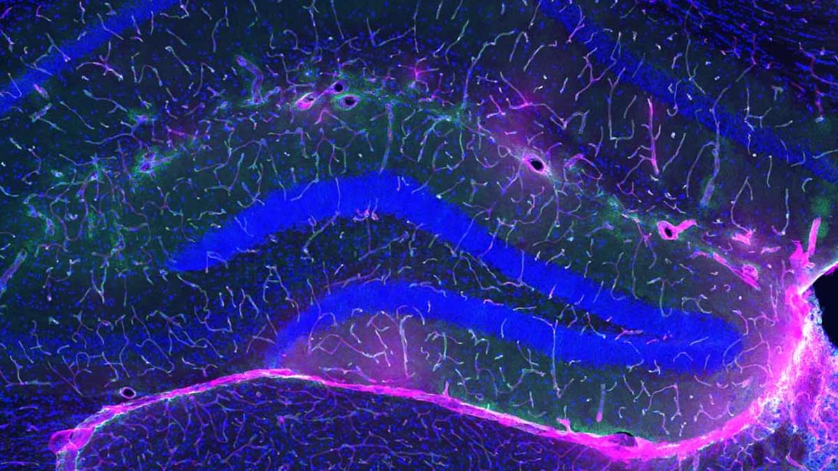
Brain vasculature in ischemic stroke.

Ischemic stroke induces dynamic cellular structural changes in the neurovascular unit, leading to disrupted structural integrity of the blood–brain barrier, neuronal degeneration, and responsive angiogenesis coordinated by endothelial cells, reactive astrocytes, and pericytes.

After ischemic stroke, the neurovascular coupling function of the neurovascular unit is also disrupted, manifested by the metabolic dysregulation of glucose, lipid/fatty acid, and amino acids.

Neurovascular unit dynamic structural remodeling and metabolic dysfunction following ischemic stroke show cellular states and spatiotemporal heterogeneities, revealing new perspectives on ischemic stroke pathogenesis and future therapeutic strategies.

Multidimensional approach aiming to repair neurovascular unit structural disorganization and restore metabolic homeostasis with cellular and spatiotemporal precision is the optimal therapeutic strategy for ischemic stroke. sciencenewshighlights ScienceMission https://sciencemission.com/neurovascular-unit-in-ischemia


The neurovascular unit (NVU) is a multicellular system functioning to maintain healthy brain homeostasis and regulate the exchange of essential elements between the blood and the brain. Recent studies have shown that, in response to ischemic stroke (IS), the NVU undergoes dynamic structural remodeling and metabolic dysfunction, revealing new features of IS pathogenesis. Recent breakthroughs in single-cell multiomics provide emerging evidence regarding the spatiotemporal heterogeneity of NVU responses to IS. To date, clinical treatments for IS-induced brain injury remain very limited. These new studies have advanced our knowledge of the dynamic cellular and molecular changes of the NVU after IS, paving the way for new therapeutic strategies.

Exercise Protects Against Alzheimer’s, And Scientists May Finally Know Why

Among its numerous health benefits, physical activity reduces the risk of developing Alzheimer’s disease. A new study on mice now dives into the specific mechanisms and proteins that allow exercise to protect our brains.

Scientists had previously determined that physical activity increases a protein called glycosylphosphatidylinositol-specific phospholipase D1 in the blood of mice, and that this protein is associated with good brain health.

That protein – more succinctly referred to as GPLD1 – strengthens the barrier that guards the brain against all sorts of unwelcome visitors within our blood, protecting against inflammation and subsequent cognitive decline.

Abstract: Presenting a cutting-edge discovery on the mechanisms by which immune cells influence health and disease at the later stages of cerebral ischemic stroke

Here, Chuan Qin & team use complementary models in experimental ischemic stroke, showing early post-stroke stages in which microglia recruit B cells into ischemic lesions through MIF/CD74/CXCR4, while later stage post-stroke effects involve interferon signaling in B cells that drives neuroinflammation and brain injury:

The image shows B lymphocytes (Green) in mouse dura tissue colocalizing with CD31+ blood vessels (Red).


1Department of Neurology, Tongji Hospital, Tongji Medical College and State Key Laboratory for Diagnosis and Treatment of Severe Zoonotic Infectious Diseases;

2Key Laboratory of Vascular Aging, Ministry of Education, Tongji Hospital of Tongji Medical College; and.

3Hubei Key Laboratory of Neural Injury and Functional Reconstruction, Huazhong University of Science and Technology, Wuhan, Hubei, China.

Basal progenitors as drivers of neocortical expansion

Neocortical expansion driven by basal progenitors.

The emergence of indirect neurogenesis, driven by highly proliferative basal progenitors, contributed to the significant enlargement of the mammalian neocortex during brain evolution.

In recent years, several human-specific genes and enhancers have been described that differentially affect the biology of progenitor cells and potentially contribute to the increased neocortical complexity and disease-susceptibility of the human brain.

Emerging research is uncovering multiple pathways that disrupt basal progenitor biology, emphasizing these pathways’ involvement not only in classical neurogenesis-related disorders such as microcephaly but also in neurodevelopmental conditions traditionally linked to impairments in neuronal connectivity. sciencenewshighlights ScienceMission https://sciencemission.com/Basal-progenitors


The diversification and expansion of distinct progenitor cell subtypes during embryogenesis are essential to form the sophisticated brain structures present in vertebrates. In particular, the emergence of highly proliferative basal progenitors contributed to the evolutionary enlargement of the mammalian neocortex. Basal progenitors are at the center of indirect neurogenesis and can be divided into two main subtypes: the classical TBR2-positive intermediate progenitor cells and the outer radial glial cells, which are especially abundant in gyrencephalic species. While the function of some transcriptomic regulators is conserved across the mammalian clade, recent studies have identified human-specific genes and enhancers that uniquely affect progenitor biology, possibly driving the increased neocortical complexity and disease-susceptibility of the human brain.

/* */