Toggle light / dark theme

The anatomy of pain and suffering in the brain and its clinical implications

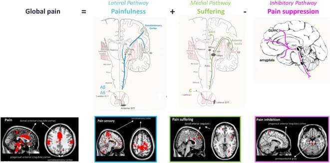
Pain is an unpleasant sensory and emotional experience associated with actual or potential tissue damage. Chronic pain, with a prevalence of 20–30% is the major cause of human suffering worldwide, because effective, specific and safe therapies have yet to be developed. It is unevenly distributed among sexes, with women experiencing more pain and suffering. Chronic pain can be anatomically and phenomenologically dissected into three separable but interacting pathways, a lateral ‘painfulness’ pathway, a medial ‘suffering’ pathway and a descending pain inhibitory pathway. One may have pain(fullness) without suffering and suffering without pain(fullness). Pain sensation leads to suffering via a cognitive, emotional and autonomic processing, and is expressed as anger, fear, frustration, anxiety and depression.

Functional photoacoustic microscopy reaches super-resolution by tracking red blood cells

The brain relies on real-time delivery of oxygen and nutrients through its microvasculature, which threads through neural tissue like electrical wires. While modern imaging technologies allow researchers to follow the activity of individual neurons in the brain, they are not yet advanced enough to dissect the microvascular function at a comparable spatial scale. This gap hinders our understanding of cerebral small vessel disease and its contributions to cognitive impairment and dementia.

To address this challenge, a team of researchers at Washington University in St. Louis and Northwestern University, led by Song Hu, professor of biomedical engineering in the McKelvey School of Engineering, have developed super-resolution functional photoacoustic microscopy (SR-fPAM).

By tracking the movement and oxygenation-dependent color change of red blood cells, SR-fPAM allows researchers to image blood flow and oxygenation at single-cell resolution in the mouse brain, which bridges a critical gap in functional microvascular imaging and could provide new insight into microvascular health and disease, such as stroke, vascular dementia and Alzheimer’s disease.

Efficient amyloid-β degradation in Alzheimer’s disease using SPYTACs

Now online! SPYTAC is a synthetic peptide-programmed targeted protein degradation platform harnessing LRP1 to drive lysosomal degradation of extracellular amyloid-β in the brain and periphery. In 5×FAD mice, SPYTAC treatment efficiently degrades amyloid-β, preserves neurons, and improves cognition with reduced neuroinflammation and microhemorrhage when compared with antibody therapy.

Eye tracking and brain signals reveal how some skills become second nature

Expertise isn’t easy to pass down. Take riding a bike: A seasoned cyclist might talk a beginner through the basics of how to sit and when to push off. But other skills, like how hard to pedal to keep balanced, are more intuitive and harder to articulate. This implicit know-how is known as tacit knowledge, and very often, it can only be learned with experience and time.

But a team of MIT engineers wondered: Could an expert’s unconscious know-how be accessed, and even taught, to quickly bring a novice up to an expert’s level?

The answer appears to be “yes,” at least for a particular type of visual-learning task.

Unbalanced chromatin binding of Polycomb complexes drives neurodevelopmental disorders

Neurodevelopmental disorders from Polycomb complex missense mutations.

The causes of many neurodevelopmental disorders (NDDs) is yet to be determined.

The researchers report new missense mutations in the Polycomb repressive complex 1 (PRC1) E3 ligases RNF2 and RING1 in individuals with neurodevelopmental disorders.

Functional dissection of a deleterious variant reveals that balanced co-recruitment of Polycomb complexes to chromatin is essential for proper neurogenesis and for normal brain function and behavior. sciencenewshighlights ScienceMission https://sciencemission.com/Polycomb-complexes-drives-NDDs


Borges, González-Blanco, Arigela, et al. report new missense mutations in the PRC1 genes RNF2 and RING1 in individuals with neurodevelopmental disorders. Functional dissection of a deleterious variant reveals that balanced co-recruitment of Polycomb complexes to chromatin is essential for proper neurogenesis and for normal brain function and behavior.

Life-changing drug identified for children with rare epilepsy

A new experimental treatment for children with a hard-to-treat form of epilepsy is safe and can reduce seizures dramatically, helping them lead much healthier and happier lives, according to the findings of a UCL (University College London) and Great Ormond Street Hospital-led international clinical trial. In a paper published in The New England Journal of Medicine, researchers found that children with Dravet syndrome had up to 91% fewer seizures while being regularly administered a new medication called zorevunersen.

The results also show, for the first time, the potential to reduce the impact of the condition on a child’s mental processes and behavior. The children’s quality of life improved over a three-year period and most of the treatment’s side effects were mild.

Dravet syndrome is a devastating genetic condition that causes frequent, hard-to-control seizures and long-term neurodevelopmental impairment. The condition also causes feeding difficulties, movement problems and has a high risk of premature death. Current treatments fail to control seizures in most patients and there are no approved medicines that address the condition’s devastating cognitive and behavioral impacts.

Restoring circadian rhythms in the hypothalamic paraventricular nucleus reverses aging biomarkers and extends lifespan in male mice

Now online! Enhancing circadian amplitude in mouse hypothalamic paraventricular nucleus neurons by 3′-deoxyadenosine treatment alleviates age-related pathologies and extends lifespan.

Ctenophore research points to earlier origins of brain-like structures

New 3D reconstructions of a key sensory organ in ctenophores reveal an unexpected structural and functional complexity. The findings suggest that an elementary brain may have already appeared in our most ancient relatives, reshaping our understanding of nervous system evolution in animals. The work is published in Science Advances.

Ctenophores (comb jellies) are gelatinous animals that appeared in the ocean an estimated 550 million years ago. The delicate animals possess a specialized sensory structure called the aboral organ (AO), which allows them to sense gravity, pressure, and light. The new morphological study reveals that this organ is far more complex than previously thought.

“We show that the AO is a complex and functionally unique sensory system,” said Pawel Burkhardt, group leader at the Michael SARS Centre, University of Bergen. “Our study profoundly enhances our understanding of the evolution of behavioral coordination in animals.”

/* */