Some of the most important tools in the toolbox of modern cell biologists are special chunks of DNA that act like spies, reporting on the cell’s function. The markers, known as reporter genes, allow researchers to get a sense for what cells are doing by watching genetic programs embedded in their DNA turn on and off.
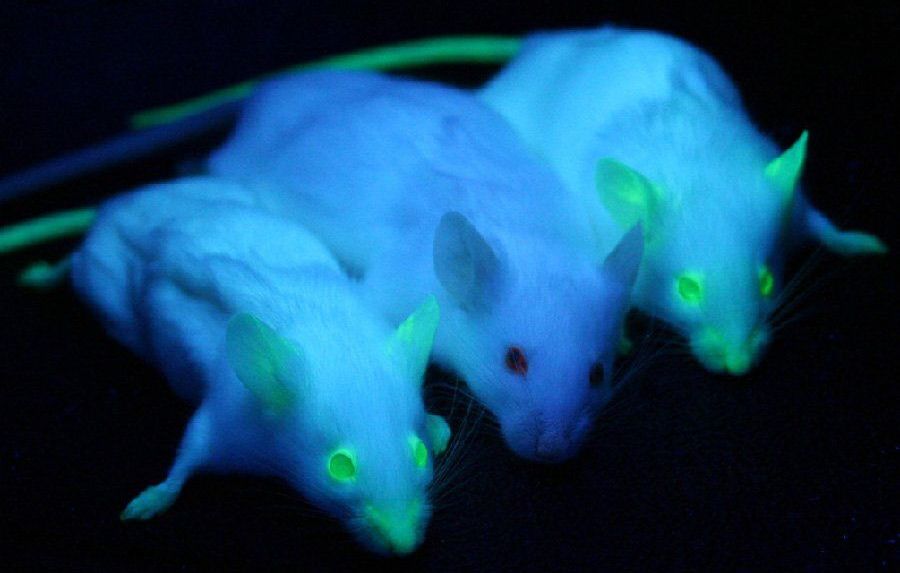
Reporter genes work by encoding proteins that can be seen from outside the cell. One particularly popular reporter gene encodes something called the green fluorescent protein (GFP), which, true to its name, is a protein that glows bright green. So, if a researcher wants to learn more about how cells become neurons, they can insert the GFP gene alongside a neuronal gene into an embryo’s DNA. When the embryo’s cells turn on the neuron gene, they will also express the GFP gene, and the cells will glow green, making it easy for the researcher to see that the genetic program that encodes neuron formation is active.
As useful as this technique has been, it has a big limitation: Because light does not penetrate well through most living tissue, the GFP gene cannot be used for monitoring the activity of cells deep inside an organism. But now, Caltech’s Mikhail Shapiro has a solution. A team consisting of Shapiro, professor of chemical engineering and investigator with the Heritage Medical Research Institute, graduate student Arash Farhadi, and their colleagues, has developed a reporter gene that allows them to see genetic activity using ultrasound, which can penetrate deeply through tissue, instead of light.