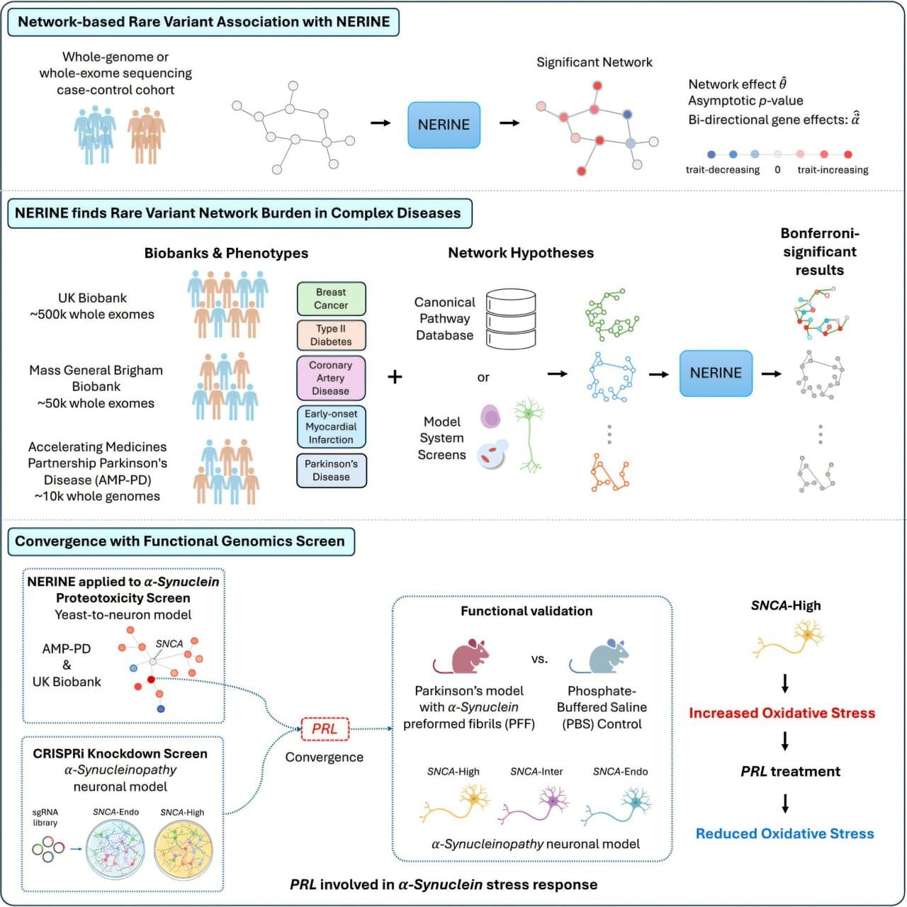
Toggle light / dark theme

Removing toxic proteins before they can damage motor neurons

University of Wollongong (UOW) scientists have developed a breakthrough therapy that clears toxic proteins from nerve cells—a discovery that advances the work of the late Professor Justin Yerbury and could transform the treatment of motor neuron disease (MND).

The proof-of-concept study, published in Nature Communications and led by Dr. Christen Chisholm from UOW’s Molecular Horizons, unveils a therapeutic designer molecule, MisfoldUbL, that targets and removes toxic misfolded SOD1 (superoxide dismutase 1) proteins from cells. SOD1 is an antioxidant enzyme that plays a crucial role in protecting cells from damage caused by superoxide radicals. About 35% of people with inherited MND in Australia have SOD1 gene mutations that cause more frequent misfolding.

“In MND, proteins misfold more frequently and the cell’s degradation systems become overwhelmed and stop working properly. The misfolded can then accumulate, forming clumps or ‘aggregates’ and over time, this accumulation damages and eventually kills motor neurons, leading to gradual muscle weakness, paralysis and death,” Dr. Chisholm said.

Phages with fully-synthetic DNA can be edited gene by gene

A team led by University of Pittsburgh’s Graham Hatfull has developed a method to construct bacteriophages with entirely synthetic genetic material, allowing researchers to add and subtract genes at will. The findings open the field to new pathways for understanding how these bacteria-killing viruses work, and for potential therapy of bacterial infections.

Sperm molecules can predict IVF success

The sperm is not a passive supplier of genetic material to the egg. A study from Linköping University, Sweden, shows that certain molecules that come with the sperm, so-called micro-RNA, contribute to the development of the embryo several days after conception. The findings, published in the journal Nature Communications, may in the long term, contribute to better diagnosis and treatment of involuntary childlessness.

“It seems that sperms can help with embryo development by bringing other molecules with them, in addition to DNA. These molecules aid in starting embryo development. So you can say that the sperm, or the male part of conception, has a greater significance than was previously understood,” says Anita Öst, professor of cell and at Linköping University, who led the study.

Many couples are affected by involuntary childlessness, or infertility. About one in six people suffer from infertility. For some, it is possible to become pregnant through what is known as in vitro fertilization, IVF, which takes place outside the body. The fertilized eggs are then transferred to the uterus and hopefully lead to pregnancy. Embryo quality is one of the major limiting factors for successful IVF treatment. Improved early embryo quality assessment could increase chances that IVF treatment leads to pregnancy.

Congenital heart disease mutation linked to kidney damage

Biomedical engineers at Duke University have shown that a genetic mutation that causes congenital heart disease also contributes to kidney damage and developmental defects. Identifying this early cause of kidney damage could enable clinicians to diagnose and address kidney problems much sooner than current practices allow. The research was published on November 3 in the journal Nature Biomedical Engineering.

Congenital heart disease (CHD) is a common cause of death in childhood and affects 1 out of every 1,000 births. The disease occurs when the heart doesn’t form correctly before birth, causing leaky valves, defective vessels, or holes in the heart. While some cases of CHD can be remedied, children with life-threatening complications often require surgery or even a heart transplant. More than 25% of patients also end up developing problems with other organs, which severely compromise life expectancy.

“Research has shown that children diagnosed with CHD almost always have kidney problems by age 4,” said Samira Musah, the Alfred M. Hunt Faculty Scholar Assistant Professor of Biomedical Engineering and Assistant Professor of Medicine at Duke University, and the senior author of the study. “Given the shared developmental origin of the heart and kidney, I wondered if a genetic mutation tied to CHD also causes the observed in affected patients.”

Mutations Lurking in Alternative Proteins May Cause Disease

Although the genetic cause of many diseases have been identified, it’s estimated that as many as 70% of patients with a rare disorder do not know what causes their disease. Millions of people live with rare diseases, and scientists are still searching for the answers to these medical mysteries. Now researchers have developed a different method for analyzing patient genetic data, which may provide clues. These findings, which were reported in Molecular Cell, have highlighted that multiple proteins can often be produced from one gene; the cell can simply interpret the sequence in different ways.

In a basic genetics lesson, a student will learn that proteins are encoded by genes, and that different genes make different proteins. But in reality, the same gene sequence may encode for multiple proteins, and it can be up to the molecular machinery of the cell to decide which of those gene sequences ends up transcribed into a protein. In fact, most genes can code for more than one protein.

Diet Composition That Corresponds To A 17y Younger Biological Age (Test #6 In 2025 Deep Dive)

Join us on Patreon! https://www.patreon.com/MichaelLustgartenPhD

Discount Links/Affiliates:
Blood testing (where I get the majority of my labs): https://www.ultalabtests.com/partners/michaellustgarten.

At-Home Metabolomics: https://www.iollo.com?ref=michael-lustgarten.
Use Code: CONQUERAGING At Checkout.

Clearly Filtered Water Filter: https://get.aspr.app/SHoPY

Epigenetic, Telomere Testing: https://trudiagnostic.com/?irclickid=U-s3Ii2r7xyIU-LSYLyQdQ6…M0&irgwc=1
Use Code: CONQUERAGING

NAD+ Quantification: https://www.jinfiniti.com/intracellular-nad-test/

Can brainless animals think?

Creatures like sea stars, jellyfish, sea urchins and sea anemones don’t have brains, yet they can capture prey, sense danger and react to their surroundings.

So does that mean brainless animals can think?

“Brainless does not necessarily mean neuron-less,” Simon Sprecher, a professor of neurobiology at the University of Fribourg in Switzerland, told Live Science in an email. Apart from marine sponges and the blob-like placozoans, all animals have neurons, he said.

Creatures like jellyfish, sea anemones and hydras possess diffuse nerve nets — webs of interconnected neurons distributed throughout the body and tentacles, said Tamar Lotan, head of the Cnidarian Developmental Biology and Molecular Ecology Lab at the University of Haifa in Israel.

“The nerve net can process sensory input and generate organized motor responses (e.g., swimming, contraction, feeding, and stinging), effectively performing information integration without a brain,” she told Live Science in an email.

This simple setup can support surprisingly advanced behavior. Sprecher’s team showed that the starlet sea anemone (Nematostella vectensis) can form associative memories — learning to link two unrelated stimuli. In the experiment, the researchers trained sea anemones to associate a harmless flash of light with a mild shock. Eventually, the light alone made them retract.

Another experiment showed that sea anemones can learn to recognize genetically identical neighbors after repeated encounters and curb their usual territorial aggression. The fact that anemones change their behavior toward genetically identical neighbors suggests they can distinguish between “self” and “non-self”

Scientists discover creature with “all-body brain”

This is the conclusion of an international team of researchers, who found that this nervous system has a genetic organization resembling that of the brain of vertebrates, like humans.

“Our results show that animals without a conventional central nervous system can still develop a brainlike organization,” said paper author and biologist Jack Ullrich-Lüter of the Natural History Museum, Berlin, in a statement.

He added: “This fundamentally changes how we think about the evolution of complex nervous systems.”

Cellular senescence and cell therapy in cardiovascular diseases

Hayflick and Moorhead initially defined cellular senescence in 1961 [10]. As senescent cells become enlarged with a flattened morphology, they exhibit an irreversible loss of proliferative potential. Changes in the expression of genetic profiles in these cells result in the secretion of pro-inflammatory molecules [11]. Senescent cells accumulate in various tissues and organs associated with aging and age-related disorders, and they are believed to become pathogenic by introducing chronic inflammation and tissue remodeling. Two major senescence-associated pathways have been highlighted in the recent literature. Telomeres are situated at both ends of a chromosome and replicate incompletely during cell division, leading to telomere shortening. When telomere shortening goes beyond the physiological range, it is recognized as DNA damage and activates replicative cellular senescence, primarily through the p53 or p16INK4a signaling pathways. p16INK4a also plays a crucial role in the mitotic process. It regulates the G1/S-phase transition of the cell cycle, helping to maintain the accuracy of cell proliferation. Normal cell division requires smooth progression through the cell cycle, and p16INK4a ensures that cells halt proliferation in the presence of DNA damage or unfavorable division conditions, thereby preserving genomic stability and preventing errors or malignancies during mitosis. Another form of cellular senescence is stress-induced premature senescence, triggered by various external and internal stress signals, including oxidative stress, irradiation, oncogenic activation, and metabolic stress. Research indicates that p53/p21 and p16INK4a signaling are primarily activated in response to DNA damage and telomere dysfunction. In contrast, p16INK4a signaling is mainly associated with mitogenic and general cellular stress [12, 13]. IGFBP7 is a member of the IGFBP family. It is a stress-responsive gene that can be upregulated in response to oxidative stress and DNA damage. The IGFBP7–p53 pathway is a critical stress–senescence pathway essential for regulating cell fate, such as cell cycle arrest, senescence, and apoptosis. This pathway may be a target for anti-tumor and anti-fibrotic therapies; however, its inhibitory effect on tissue regeneration should also be considered [14]. Senescent cells exhibit various morphological and biochemical characteristics that aid their detection [15]. Currently, no single marker can definitively identify a senescent cell; instead, combinations of markers and analytical techniques are commonly employed to improve detection specificity. Table 1 displays some widely used markers for this purpose. Many stressors that induce senescence activate the p53/p21 or p16INK4a pathways. However, it’s important to note that activating these signaling pathways does not provide conclusive evidence that the cells are senescent [16]. Currently, senescence-associated ß-galactosidase (SA-ß-galactosidase) is widely used to identify senescent cells as a marker of senescence, which has a pH optimum of 6.0; however, the SA-ß-gal activity is also known to increase in fibroblasts cultured under serum starvation [17,18,19]. Another category of sensitive senescence indicators includes DNA damage response (DDR) gene products, which are usually visualized through immunofluorescence. The DDR protein most commonly used for this purpose is γH2AX phosphorylated at Ser-139, which accumulates at sites of double-stranded DNA breaks and facilitates the detection of proteins involved in the double-strand break repair pathway [20, 21]. DNA damage at telomeres suggests that both cardiomyocytes and various non-cardiomyocytes, including myofibroblasts, endothelial cells, and vascular smooth muscle cells, contribute to the senescence of the cardiovascular system. These cells interact within the microenvironment, altering cardiovascular function and promoting disease progression. Additionally, some studies have monitored cytokine secretion related to the senescence-associated secretory phenotype (SASP), characterized by the extensive release of pro-inflammatory compounds. Common SASP factors secreted by senescent cells include signaling molecules such as interleukins (IL-6, IL-1ß, IL-8) and other factors [22, 23]. The cell makers mentioned above are all related to senescence, but do not exist in isolation.

In summary, cells that show positive senescent markers are well recognized for their causal roles in the progression of pathologies associated with age-related diseases [24, 25]. Investigating biological markers that provide direct evidence of cellular senescence continues to be a significant area of research. In this review article, we aim to outline the role of senescence in cardiovascular disease and explore the potential of therapies targeting senescent cells.

Cardiomyocytes comprise 25–35% of the total number of cells in the heart [26]. Their cell cycle arrest cannot easily define the senescence of cardiomyocytes because they are terminally differentiated cells. Cardiomyocytes undergo cell cycle arrest due to the activation of the DNA damage response triggered by exposure to higher oxygen concentrations in the postnatal environment [27]. The accumulating environment indicates these cells retain proliferative capacity. It was reported that cardiomyocyte turnover was < 1% per year [28]. Senescent cardiomyocytes exhibit significant functional, morphological, and metabolic differences compared to normal cardiomyocytes. Hallmarks of senescent cardiomyocytes include mitochondrial dysfunction, DNA damage, contractile dysfunction, endoplasmic reticulum (ER) stress, SASP, and hypertrophic growth [29].

/* */