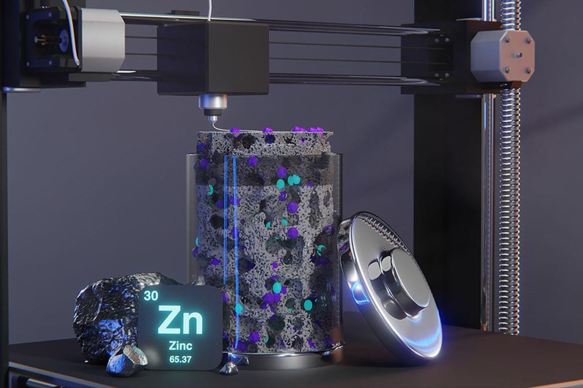
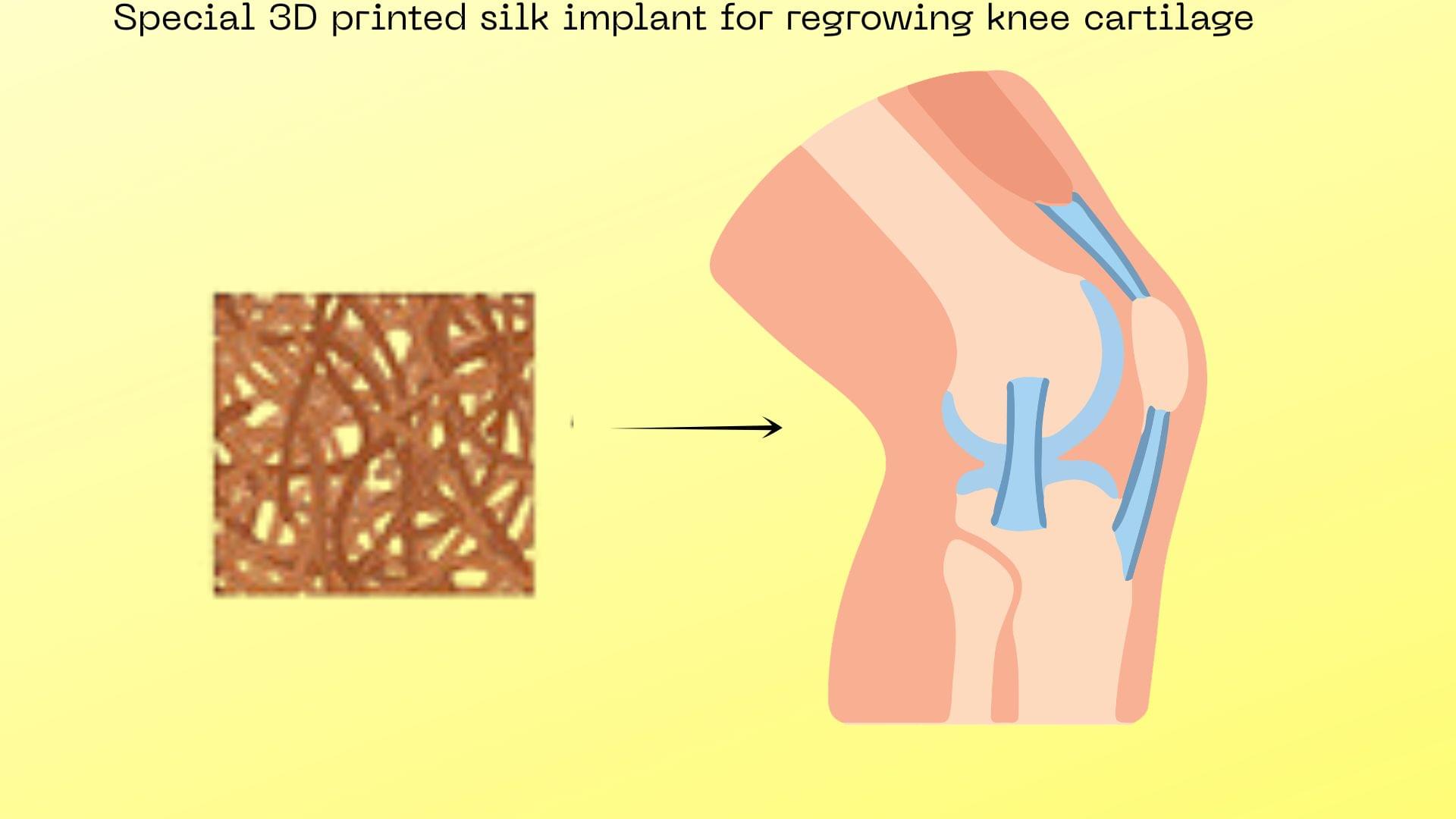
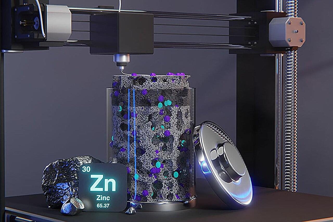
Toggle light / dark theme

AI-driven research is making titanium 3D-printing faster, stronger, and more efficient, transforming aerospace and defense manufacturing.

Producing high-performance titanium alloy.

A mixture of two metallic elements typically used to give greater strength or higher resistance to corrosion.

Empa researchers are working on artificial muscles that can keep up with the real thing. They have now developed a method of producing the soft and elastic, yet powerful structures using 3D printing. One day, these could be used in medicine or robotics – and anywhere else where things need to move at the touch of a button.


A team of researchers from Empa’s Laboratory for Functional Polymers is working on actuators made of soft materials. Now, for the first time, they have developed a method for producing such complex components using a 3D printer. The so-called dielectric elastic actuators (DEA) consist of two different silicone-based materials: a conductive electrode material and a non-conductive dielectric. These materials interlock in layers. “It’s a bit like interlacing your fingers,” explains Empa researcher Patrick Danner. If an electrical voltage is applied to the electrodes, the actuator contracts like a muscle. When the voltage is switched off, it relaxes to its original position.

3D printing such a structure is not trivial, Danner knows. Despite their very different electrical properties, the two soft materials should behave very similarly during the printing process. They should not mix but must still hold together in the finished actuator. The printed “muscles” must be as soft as possible so that an electrical stimulus can cause the required deformation. Added to this are the requirements that all 3D printable materials must fulfill: They must liquefy under pressure so that they can be extruded out of the printer nozzle. Immediately thereafter, however, they should be viscous enough to retain the printed shape. “These properties are often in direct contradiction,” says Danner. “If you optimize one of them, three others change … usually for the worse.”

Despite its success, FNP has some limitations: it can’t create stable particles larger than 400 nm, the maximum drug content is about 70 percent, the output is low, and it can only work with very hydrophobic (water-repelling) molecules. These issues arise because the particle core formation and particle stabilization happen simultaneously in FNP. The new SNaP process overcomes these limitations by separating the core formation and stabilization steps.

In the SNaP process, there are two mixing steps. First, the core components are mixed with water to start forming the particle core. Then, a stabilizing agent is added to stop the core growth and stabilize the particles. This second step must happen quickly, less than a few milliseconds after the first step, to control the particle size and prevent aggregation. Current SNaP setups connect two specialized mixers in series, controlling the delay time between steps. However, these setups face challenges, including high costs and difficulties in achieving short delay times needed for small particle formation.

A new approach using 3D printing has solved many of these challenges. Advances in 3D printing technology now allow the creation of precise, narrow channels needed for these mixers. The new design eliminates the need for external tubing between steps, allowing for shorter delay times and preventing leaks. The innovative stacked mixer design combines two mixers into a single setup, making the process more efficient and user-friendly.

An international collaboration headed by researchers in the Department of Physics has shown that additive manufacturing offers a realistic way to build large-scale plastic scintillator detectors for particle physics experiments.

In 2024, the T2K Collaboration started to collect new neutrino data following several upgrades to the experiment that included new types of detectors. One of these, called SuperFGD, has a mass of about 2 tons of sensitive volume and is made of approximately two million cubes. Each cube is made of plastic scintillator (PS) material that emits light when a charged particle passes through it.

Neutrinos carry no charge, as their name indicates, but they sometimes interact with other particles, then produce electrons, protons, muons or pions that can be detected. Each PS cube is traversed by three orthogonal optical fibers that collect the scintillation light and guide it to 56,000 photodetectors. The data reveal three-dimensional (3D) particle tracks, which in turn allow researchers to learn more about neutrinos.

Charging forward at top speed, a garden snail slimes up 1 millimeter of pavement per second. By this logic, Beckman Institute for Advanced Science and Technology researchers’ new 3D printing process speeds past existing methods—at a snail’s pace.

Researchers in Beckman’s Autonomous Materials Systems Group created “growth printing,” which mimics tree trunks’ outward expansion to print polymer parts quickly and efficiently without the molds and expensive equipment typically associated with 3D printing. Their work appears in the journal Advanced Materials.

“Humans are incredibly talented at making things. Completely new manufacturing processes are hard to find. Growth printing is entirely new, which is thrilling,” said Sameh Tawfick, a professor of mechanical science and engineering at the University of Illinois Urbana-Champaign and project lead.

Empa researchers are working on producing artificial muscles that can keep up with the real thing. They have now developed a method of producing the soft and elastic yet powerful structures using 3D printing.

One day, these could be used in medicine or robotics—and anywhere else where things need to move at the touch of a button. The work is published in the journal Advanced Materials Technologies.

Artificial muscles don’t just get robots moving: One day, they could support people at work or when walking, or replace injured muscle tissue. However, developing artificial muscles that can compare to the real thing is a major technical challenge.

Marine worms use nature’s 3D printing to build their bristles piece by piece.


A new study has shed light on how certain marine worms form bristles, hair-like projections on each side.

A team of researchers, led by molecular biologist Florian Raible from the Max Perutz Labs at the University of Vienna, used advanced imaging techniques to closely study Platynereis dumerilii, which is often considered a living fossil.

These annelid worms have extraordinary bristles that enable them to navigate their aquatic environment. But how are these intricate structures formed? It turns out that these species develop bristles piece by piece, similar to the process of 3D printing.

3D printing is revolutionizing microbial electrochemical systems (MES) by enabling precise reactor design, custom electrode fabrication, and enhanced bioprinting applications. These innovations optimize pollutant degradation and energy production, with significant implications for sustainability and environmental management.

Microbial electrochemical systems (MES) are emerging as a promising technology for addressing environmental challenges by leveraging microorganisms to transfer electrons. These systems can simultaneously degrade pollutants and generate electricity, making them valuable for sustainable wastewater treatment and energy production.

However, conventional methods for constructing MES components often lack design flexibility, limiting performance optimization. To overcome these limitations and enhance MES efficiency, innovative fabrication techniques are needed—ones that allow precise control over reactor structures and functions.

Biomedical engineers at the University of Melbourne have developed a 3D bioprinting system capable of creating structures that closely replicate various human tissues, ranging from soft brain tissue to more rigid materials like cartilage and bone.

This innovative technology provides cancer researchers with a powerful tool for replicating specific organs and tissues, enhancing their ability to predict drug responses and develop new treatments. By offering a more accurate and ethical approach to drug discovery, it also has the potential to reduce reliance on animal testing.

Head of the Collins BioMicrosystems Laboratory at the University of Melbourne, Associate Professor David Collins said: In addition to drastically improving print speed, our approach enables a degree of cell positioning within printed tissues. Incorrect cell positioning is a big reason most 3D bioprinters fail to produce structures that accurately represent human tissue.