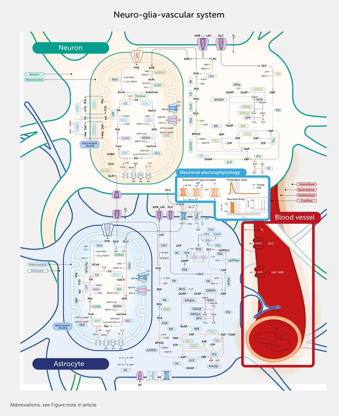
Toggle light / dark theme

Aging human breast atlas reveals cancer susceptibility

The team used advanced imagining techniques to analyse breast tissue from more than 500 women aged 15 to 86 years old. The tissue included biopsies taken from women for non-cancer-related reasons.

Combining these images with details of the hormone receptors and immune cells present, as well as the tissue architecture, the researchers were able to map how breast tissue changes over time in unprecedented detail. Their findings point to reasons why breast cancer risk increases with age and why tumors in younger women differ biologically.

The author added: “Our map revealed that as women age, their breast tissue goes through major changes, with the most dramatic changes occurring at menopause. There are changes, too, during their twenties, possibly linked to pregnancy and childbirth, but these are far less pronounced.”

The map revealed that all types of cells become fewer in number and divide far less often. Milk-producing structures known as lobules shrink or disappear, while the ducts that that carry milk become relatively more common, with the supporting layer around them becoming thicker. Fat cells increase while blood vessels decrease.

Meanwhile, changes occur in the immune environment. Younger breasts have more B cells and active T cells, which helps them identify and kill cancer cells. As tissue ages, these types of cells decline in number, replaced by other types of immune cell that indicate a more inflammatory and potentially less protective immune environment. ScienceMission sciencenewshighlights.


Scientists have created the most detailed map to date, comprised of over 3 million cells, showing how breast tissue changes as women age – including dramatic changes during menopause.

Tau mutation drives autophagy-lysosome dysfunction

The researchers studied a specific mutation in a brain protein called tau that causes the protein to become misfolded and alter its normal function. In general, when tau proteins become misfolded, they build up inside neurons and contribute to various forms of dementia, including Alzheimer’s dementia and frontotemporal dementia, a neurodegenerative disease similar to Alzheimer’s that often strikes earlier — in middle age — and typically involves significant changes in personality and behavior that precede cognitive decline.

In this new study, the researchers studied neurons that had been reprogrammed from skin cells sampled from patients with frontotemporal dementia who carried the tau mutation. In the neurons, the mutated tau proteins caused waste-recycling centers called lysosomes, which are involved in autophagy, to become dysfunctional, allowing cellular waste to accumulate in the lysosomes, which may contribute to neuronal death. The researchers found that enhancing autophagy with an analog of the chemical compound G2 improved clearance of the garbage, reduced tau levels in the lysosomes and prevented cellular toxicity and death.

G2 was discovered in 2019 via screening experiments seeking drugs that could reduce the accumulation of an aggregation-prone protein in a C. elegans model of alpha-1-antitrypsin deficiency, which can cause severe liver disease. The compound was later shown to boost autophagy function in mammalian cell model systems.

The researchers also have shown that G2 can protect brain cells from death in cells modeling Huntington’s disease, a fatal inherited neurodegenerative disease caused by a genetic mutation present at birth. In the cellular model of Huntington’s disease, the compound prevented the buildup of a harmful RNA molecule. ScienceMission sciencenewshighlights.


New research adds to growing evidence that helping brain cells break down and eliminate their own cellular waste is a promising treatment strategy for a variety of neurodegenerative diseases. In lab experiments, the researchers found that exposure to a novel compound can clear a harmful protein from human neurons modeling frontotemporal dementia — a devastating and ultimately fatal condition — and prevent those neurons from dying.

The study is published in the journal Nature Communications.

Hydroxyl radicals in UV-exposed water reveal surprising reaction pathway

How do radicals form in aqueous solutions when exposed to UV light? This question is important for health research and environmental protection. For example, with regard to the overfertilization of water bodies by intensive agriculture. A team at BESSY II has now developed a new method of investigating hydroxyl radicals in solution. By using a clever trick, the scientists gained surprising insights into the reaction pathway. The findings are published in the Journal of the American Chemical Society.

Hydroxyl radicals (OH·) are found everywhere, from the troposphere to the cells of the human body. There, they cause oxidative stress and accelerate the aging process. They are also increasingly present in rivers and lakes, where they are formed by the photolysis of nitrogen oxides that have entered the water from over-fertilized soils. When UV radiation from sunlight strikes nitrogen oxides, hydroxyl radicals and a range of other radicals are generated. The chemistry of these radicals is extremely difficult to characterize accurately, as they react very quickly.

A team led by Professor Alexander Föhlisch of the HZB has investigated the chemistry of hydroxyl radicals formed from nitrogen oxides in water using X-ray absorption spectroscopy at the BESSY II X-ray source.

Double‐Pronged NAD Preservation: Delaying Cellular Senescence and Initiating Musculoskeletal Regeneration

A novel synergistic drug combination (N + A) consisting of an NAD+ precursor (NMN) and an NAD+ consumption (CD38) inhibitor (API) promotes musculoskeletal regeneration in aging. Notably, increased NAD+ serves as a coenzyme for SIRT3, exerting a robust anti-senescence effect, thus promoting tri-lineage differentiation into chondrocytes, osteoblasts, and myocytes. Furthermore, oral administration of the N + A formulation modulated the intestinal microenvironment, promoting the gut microbiota-derived production of the metabolite PHS, thereby exerting indirect anti-aging effects in musculoskeletal disorders.

Key protein found to protect cartilage, offering new hope for osteoarthritis treatment

Osteoarthritis, a condition that causes pain and reduced mobility in joints such as the knees and fingers, is one of the most common joint disorders worldwide, particularly among aging populations. The disease is characterized by the gradual breakdown of cartilage, which normally cushions the bones within joints.

Despite its prevalence, current treatments for osteoarthritis mainly focus on alleviating pain rather than addressing the underlying cause of cartilage degeneration. Effective therapies that can halt or reverse cartilage damage remain limited.

A joint research team led by Dr. Chul-Ho Lee and Dr. Yong-Hoon Kim at the Laboratory Animal Resource Center of the Korea Research Institute of Bioscience and Biotechnology (KRIBB), in collaboration with Prof. JinHyun Kim at Chungnam National University Hospital, has identified a key protein, SHP (NR0B2), that plays a critical protective role in cartilage and may offer a new therapeutic strategy for osteoarthritis. The paper is published in the journal Nature Communications.

Histone modification clocks for robust cross-species biological age prediction and elucidating senescence regulation

Building upon these insights, we constructed 36 histone modification-based epigenetic clocks, which exhibited robust predictive accuracy (mean Pearson’s r = 0.91) across multiple tissues and marks. Among these, the blood-derived H3K27ac clock emerged as a particularly powerful model, outperforming several established DNA methylation clocks under matched conditions. This performance is remarkable considering that DNA methylation clocks have undergone extensive optimization over the past decade (9, 16, 18), while our histone-based approach represents a first-generation effort.

A distinctive advantage of our histone-based clocks is their resilience to technical and biological noise. When exposed to artificial Gaussian noise, the histone-based clock maintained stable predictive performance, in contrast to the sharp degradation observed in many methylation-based models. This robustness is likely attributable to the broader, structural nature of histone mark signals, which may be less sensitive to local fluctuations than single CpG methylation values. This characteristic makes histone clocks potentially more suitable for noisy, heterogeneous, or clinically derived datasets where sample quality may vary.

The practical utility of our histone-based clocks was further demonstrated by their ability to detect biological age acceleration in leukemia samples and capture age reversal following therapeutic interventions. These applications highlight the potential of histone-based clocks as biomarkers for disease states and treatment responses, offering a complementary approach to existing clinical tools.

APOE4 Increases Neurons’ Excitability Before Symptoms Appear

The pro-Alzheimer’s allele APOE4 makes hippocampal neurons in mice smaller and hyperexcitable. This effect, which resembles epilepsy and accelerated aging, can be mitigated by manipulating a neuronal protein [1].

Before symptoms arise

Alzheimer’s disease begins long before symptoms appear, building silently for decades. The single strongest genetic risk factor for the common, late-onset form of Alzheimer’s is the ε4 variant of the apolipoprotein (APOE) gene, APOE4. Carrying a single copy of this variant (being heterozygous) roughly triples your Alzheimer’s risk; having two copies increases it about 12-fold.

How an Alzheimer’s Risk Gene Disrupts Brain Circuits Long Before Memory Loss

Researchers at the Gladstone Institute have uncovered the molecular mechanism by which APOE4 — the most significant genetic risk factor for Alzheimer’s disease, present in roughly a quarter of the population — begins damaging neural circuits well before any cognitive symptoms emerge. Studying young mice carrying the APOE4 variant, the team found that the gene triggers overproduction of the protein Nell2, which causes neurons to shrink and become hyperactive. Crucially, the degree of early neuronal hyperactivity predicted the severity of memory impairment later in life, even in animals that still showed normal learning and memory at the time of measurement. Strikingly, targeting Nell2 therapeutically was able to reverse these changes even in adult animals, demonstrating that the neurodegeneration is not irreversible and that a window for intervention may exist even after the disease process has begun. The team is currently continuing preclinical testing of this therapeutic strategy.


New findings on the APOE4 gene variant point to a potential therapeutic target for Alzheimer’s disease. From left to right, Gladstone scientists Misha Zilberter, Yadong Huang, and Dennis Tabuena examine findings from their research, which is published in the journal Nature Aging.

For the millions of people who carry the gene APOE4, the strongest known genetic risk factor for Alzheimer’s disease, their brain activity may begin changing long before any memory problems appear. Now, researchers at Gladstone Institutes have uncovered a precise chain of molecular events behind those early changes and identified a potential way to reverse them.

Published in the journal Nature Aging, their new study in mouse models reveals how APOE4 triggers increased production of the protein Nell2, which makes neurons shrink and become hyperactive. The more hyperactive the neurons were in early life, the more severe were the memory problems the mice developed later in life.

/* */