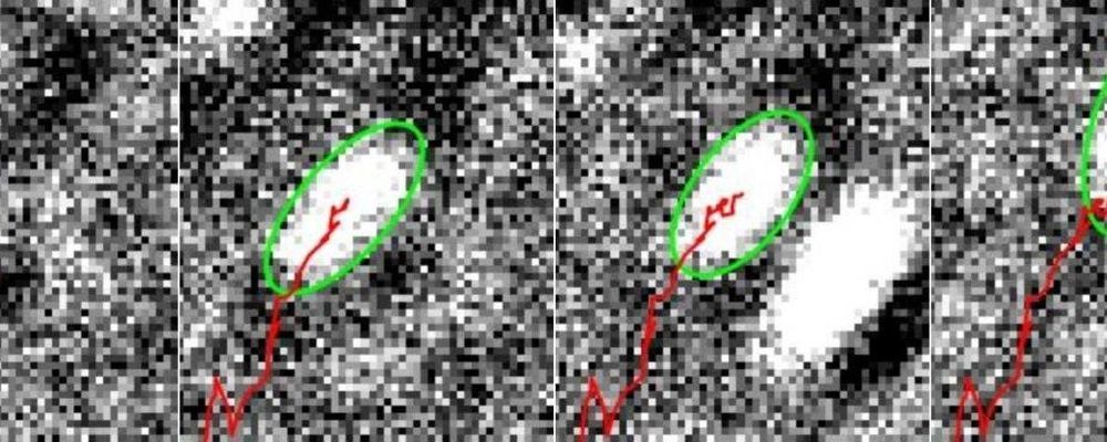
The movements of cell muscles in the form of tiny filaments of proteins have been visualised at unprecedented detail by University of Warwick scientists.
In a study published in the Biophysical Journal, scientists from the University’s Department of Physics and Warwick Medical School have used a new microscopy technique to analyse the molecular motors inside cells that allow them to move and reshape themselves, potentially providing new insights that could inform the development of new smart materials.
Myosin is a protein that forms the motor filaments that give a cell stability and are involved in remodelling the actin cortex inside the cell. The actin cortex is much like the backbone of the cell and gives it its shape, while the myosin filaments are similar to muscles. By ‘flexing’, they enable the cell to exert forces outside of it and to propagate.
