New methods enable scientists at IST Austria to take a look at the innermost of cells: High-resolution images of deep-frozen cells show structures that previously could only be guessed at.
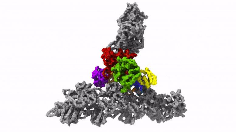
The cells in our body are in motion. Some migrate from A to B to heal wounds or fight pathogens. They do so with the help of small “feet” at the leading edge of migrating cells, so-called lamellipodia. These thin extensions are pushed forward and bind to the surface while the rest of the cell is pulled along. Inside these feet is a dense network of interwoven protein threads, called actin filaments, which form the cell’s cytoskeleton. So far, it was unclear how the Arp2/3 complex, an assembly of seven proteins central for cell motility, sprouts off new actin filaments from pre-existing ones and thus generates dense, branched networks providing the required protrusive forces to the cell.
Until now, scientists had to decide when they wanted to analyze the structure of the Arp2/3 complex: One option was to study it in isolation, where the protein complex is in an inactive conformation and hence does not allow understanding of how the network is formed. In order to become fully activated, however, the Arp2/3 complex needs to be bound to actin filaments. This requires using a method called electron tomography, which comes at the cost of considerably lower resolution. “Previous electron tomography data of Arp2/3 complexes bound to actin filaments in a test-tube environment was too imprecise, making it impossible to unambiguously tell where the individual elements of the complex must be located,” explains Florian Fäßler, a postdoc in the group of IST Austria professor Florian Schur.