It’s been a truth for a long time: if you want to study the movement and behavior of single atoms, electron microscopy can give you what X-rays can’t. X-rays are good at penetrating into samples—they allow you to see what happens inside batteries as they charge and discharge, for example—but historically they have not been able to spatially image with the same precision electrons can.
But scientists are working to improve the image resolution of X-ray techniques. One such method is X-ray tomography, which enables non-invasive imaging of the inside of materials. If you want to map the intricacies of a microcircuit, for example, or trace the neurons in a brain without destroying the material you are looking at, you need X-ray tomography, and the better the resolution, the smaller the phenomena you can trace with the X-ray beam.
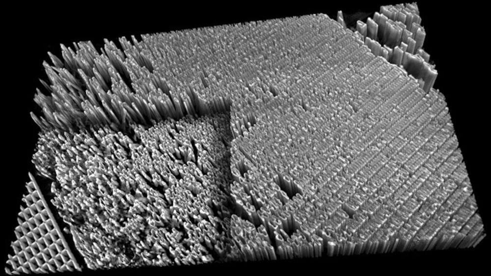
To that end, a group of scientists led by the U.S. Department of Energy’s (DOE) Argonne National Laboratory has created a new method for improving the resolution of hard X-ray nanotomography. (Nanotomography is X-ray imaging on the scale of nanometers. For comparison, an average human hair is 100,000 nanometers wide.) The team constructed a high-resolution X-ray microscope using the powerful X-ray beams of the Advanced Photon Source (APS) and created new computer algorithms to compensate for issues encountered at tiny scales. Using this method, the team achieved a resolution below 10 nanometers.