Research by Dr. Silvia de Santis and Dr. Santiago Canals, both from the Institute of Neurosciences UMH-CSIC (Alicante, Spain), has made it possible to visualize for the first time and in great detail brain inflammation using diffusion-weighted Magnetic Resonance Imaging. This detailed “X-ray” of inflammation cannot be obtained with conventional MRI, but requires data acquisition sequences and special mathematical models. Once the method was developed, the researchers were able to quantify the alterations in the morphology of the different cell populations involved in the inflammatory process in the brain.
An innovative strategy developed by the researchers has made possible this important breakthrough, which is published today in the journal Science Advances and which may be crucial to change the course of the study and treatment of neurodegenerative diseases.
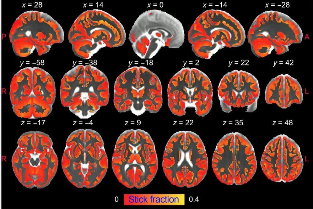
The research demonstrates that diffusion-weighted MRI can noninvasively and differentially detect the activation of microglia and astrocytes, two types of brain cells that are at the basis of neuroinflammation and its progression.