The iconic X-shaped organization of metaphase chromosomes is frequently presented in textbooks and other media. The drawings explain in captivating manner that the majority of genetic information is stored in chromosomes, which transmit it to the next generation. “These presentations suggest that the chromosome ultrastructure is well-understood. However, this is not the case,” says Dr. Veit Schubert from IPK’s chromosome structure and function research group.
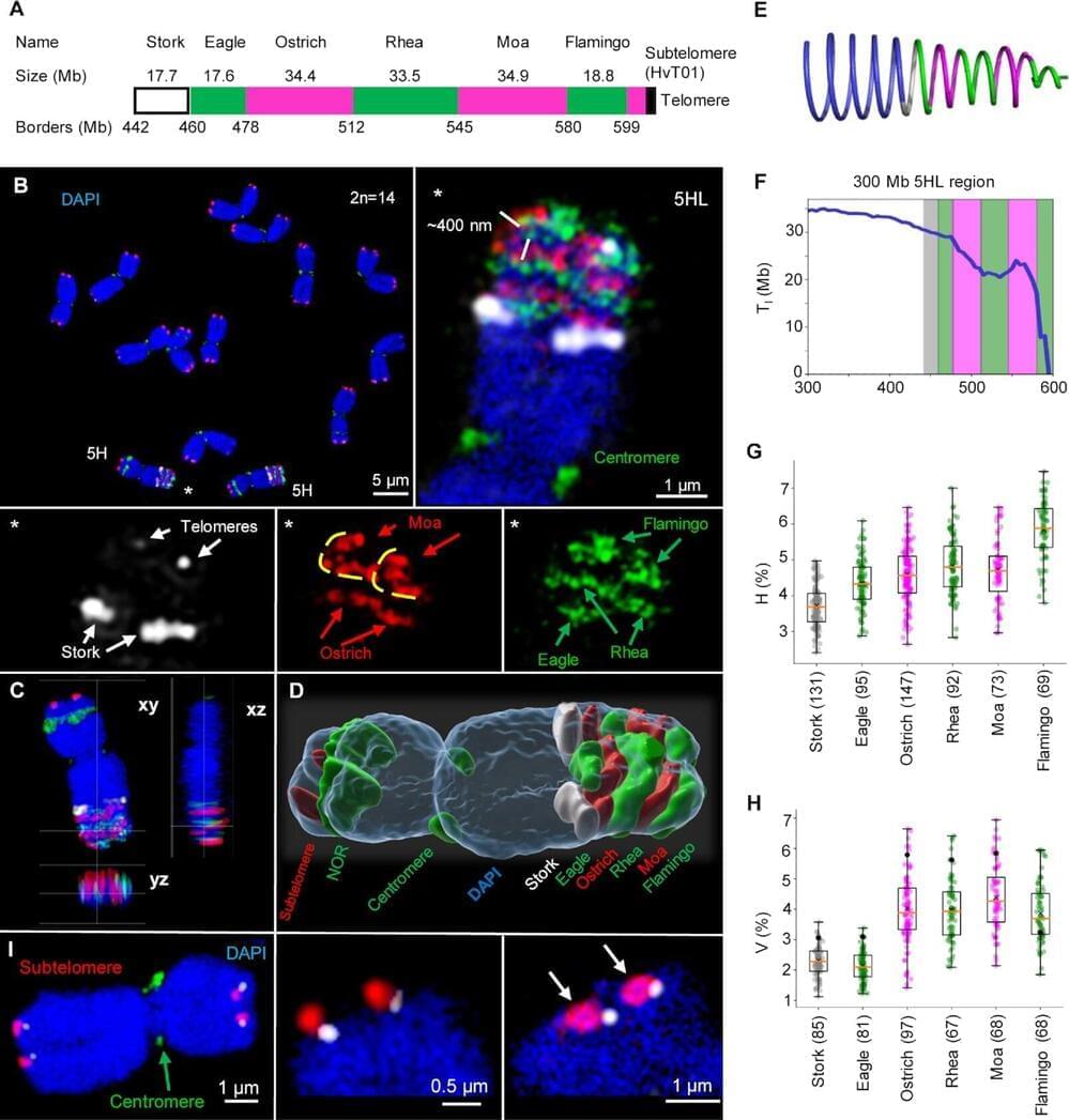
Several models have been proposed to describe the higher-order structure of metaphase chromosomes based on data obtained using a range of molecular and microscopy methods. These models are categorized as helical and non-helical. Helical models assume that the chromatin in each sister chromatid at metaphase is arranged as a coil, whereas non-helical models suggest that chromatin is folded within the chromatids without forming a spiral.
The researchers revived the term “chromonema,” which was used for the first time at the beginning of the 20th century. Now, the IPK and IEB researchers provided a detailed description of its ultrastructure. Different experimental approaches, including chromosome conformation capture sequencing (Hi-C) of isolated mitotic chromosomes, polymer modeling, and microscopic observations of sister chromatid exchanges and oligo-FISH labeled regions at the super-resolution level provided an independent proof for the coiling of the chromonema.
