Human brain organoids are an intrinsically self-organized neuronal ensemble grown from three-dimensional assemblies of human-iPSCs. As shown here, brain organoids offer a window into the complex neuronal activity that emerges from intrinsically-formed circuits capable of mirroring aspects of the developing human brain32. Applying high-density CMOS MEA to large multi-cellular networks spanning millimeters of the brain organoid cross-sections we isolated single-unit activity and computed the timing of successive action potentials not due to refractoriness referred to as ISIs. As observed in neocortical neurons in vivo, we observed action potentials with irregular ISI’s that followed a Poisson-like process. From a set of 224 neurons analyzed from four different organoids, 16% ± 8% of the total units fit a Poisson distribution (Fig. 3) with, by definition, the CV approaches one for a perfectly homogenous Poisson process, whereas purely periodic distributions have CV values of zero. Thus, a minority fraction of ISIs were highly irregular (Fig. 3), whereas a majority displayed comparatively more regular spiking patterns with less variation (denoted by a lower CV), which may function to send lower-noise spike-rate signals. ISI distributions have also been fitted to gamma distributions that are mathematically equivalent to an exponential distribution when the shape parameter (k) is one and converges to a normal distribution for large k, thus providing a useful measure of ISI-regularity similar to the CV28. Depending on architectonically defined brain regions with specialized cellular compositions and intrinsic circuitry, neurons process information differently67,68,69. Indeed, neuronal firing varies considerably across cortical regions of monkeys28,70,71. Therefore, different organizational features across the brain organoid may exhibit different dynamics to account for the observed ISI distributions. The minority fraction of irregular ISI distributions may be a feature of higher levels of entropy and circuit complexity and contain increased capacity for computation and information transfer as found in prefrontal cortex compared to more regular firing patters found in motor regions28.
We derived a graph of weighted edges that couple single unit node pairs to send and receive spikes over a wide spatial range. Due to the thickness of our organoid slices, many neurons in the slice are too far from any electrode for their spikes to be detected53. Thus, we cannot rule out the possibility that intermediate undetected neurons may account for the coupling between two correlated units. The graph does not imply downstream or upstream routes of information transfer beyond the individual binary couplings. Importantly, what the network does demonstrate is a non-random pattern of a relatively small number of statistically strong (reliable) couplings against a backdrop of weaker couplings. As demonstrated in the murine brain51,52, high anatomical connection strength edges shape a non-random framework against a background of weaker ones (Fig. 6 and Supplementary Fig. 14). The majority of the singe units (nodes), which we refer to as brokers, have large proportions of incoming and outgoing edges. The dynamic balance among receivers and senders could likely reflect short-term plasticity72.
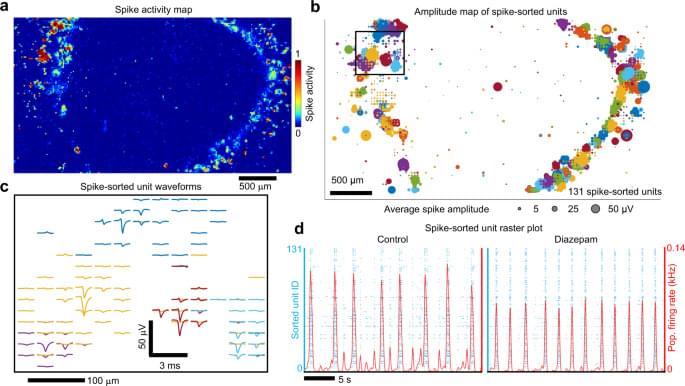
Brain organoids—composed of roughly one million cells—have neuronal assemblies of sufficient size, cellular orientation, connectivity and co-activation capable of generating field potentials in the extracellular space from their collective transmembrane currents. The basis for low frequency LFPs may be the cellular diversity that emerges in the organoid from the variety of GABAergic cells (Fig. 2), consistent with their role in the generation of highly correlated activity networks detected as LFPs31, parvalbumin cells (Fig. 2c), associated with sustaining network dynamics73, and axon tracts that extended over millimeters (Fig. 2b). Coherence of theta oscillations over spatial extents of the organoid was observed and was unlikely due to volume conduction from distant sources, as happens in EEG and MEG measurements54, because the voltage recordings were conducted within a small tissue volume (≈3.5 mm3). Consistent with minimal volume conduction effects, we validated theta oscillations by demonstrating that the imaginary part of coherency54 projected onto the same spatial locations identified by cross-correlation analysis (Supplementary Fig. 19). Correlations between theta oscillations and local neuronal firing (Fig. 7) strongly supported a local source for the rhythmic activity19,20,53. The local volume through which theta dispersed extended to the z-dimension as shown with the Neuropixels shank (Fig. 9).