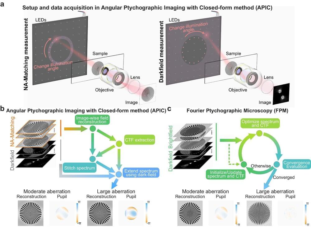
For hundreds of years, the clarity and magnification of microscopes were ultimately limited by the physical properties of their optical lenses. Microscope makers pushed those boundaries by making increasingly complicated and expensive stacks of lens elements. Still, scientists had to decide between high resolution and a small field of view on the one hand or low resolution and a large field of view on the other.
In 2013, a team of Caltech engineers introduced a microscopy technique called FPM (for Fourier ptychographic microscopy). This technology marked the advent of computational microscopy, the use of techniques that wed the sensing of conventional microscopes with computer algorithms that process detected information in new ways to create deeper, sharper images covering larger areas. FPM has since been widely adopted for its ability to acquire high-resolution images of samples while maintaining a large field of view using relatively inexpensive equipment.
Now the same lab has developed a new method that can outperform FPM in its ability to obtain images free of blurriness or distortion, even while taking fewer measurements. The new technique, described in a paper that appeared in the journal Nature Communications, could lead to advances in such areas as biomedical imaging, digital pathology, and drug screening.
