БЛОГ

Archive for the ‘bioprinting’ category: Page 4

Feb 27, 2023

3D bioprinting inside the human body could be possible thanks to new soft robot

Posted by in categories: 3D printing, bioengineering, bioprinting, biotech/medical, robotics/AI

Engineers from UNSW Sydney have developed a miniature and flexible soft robotic arm which could be used to 3D print biomaterial directly onto organs inside a person’s body.

3D bioprinting is a process whereby biomedical parts are fabricated from so-called bioink to construct natural tissue-like structures.

Continue reading “3D bioprinting inside the human body could be possible thanks to new soft robot” »

Dec 31, 2022

Microfluidic bioprinting of tough hydrogel-based vascular conduits for functional blood vessels

Posted by in categories: bioprinting, biotech/medical

Rationally designed bioinks enable bioprinting of mechanically and physiologically relevant vascular conduits.

Dec 15, 2022

Skin bioprinting: the future of burn wound reconstruction?

Posted by in categories: 3D printing, bioengineering, bioprinting, biotech/medical, robotics/AI

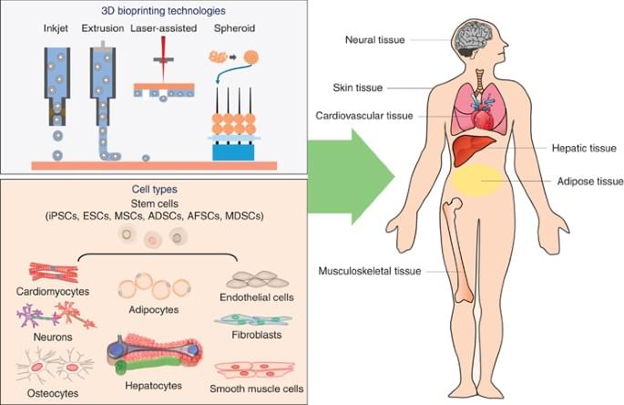
In addition to laser-assisted bioprinting, other light-based 3D bioprinting techniques include digital light processing (DLP) and two-photon polymerization (TPP)-based 3D bioprinting. DLP uses a digital micro-mirror device to project a patterned mask of ultraviolet (UV)/visible range light onto a polymer solution, which in turn results in photopolymerization of the polymer in contact [56, 57]. DLP can achieve high resolution with rapid printing speed regardless of the layer’s complexity and area. In this method of 3D bioprinting, the dynamics of the polymerization can be regulated by modulating the power of the light source, the printing rate, and the type and concentrations of the photoinitiators used. TPP, on the other hand, utilizes a focused near-infrared femtosecond laser of wavelength 800 nm to induce polymerization of the monomer solution [56]. TPP can provide a very high resolution beyond the light diffraction limit since two-photon absorption only happens in the center region of the laser focal spot where the energy is above the threshold to trigger two-photon absorption [56].

The recent development of the integrated tissue and organ printer (ITOP) by our group allows for bioprinting of human scale tissues of any shape [45]. The ITOP facilitates bioprinting with very high precision; it has a resolution of 50 μm for cells and 2 μm for scaffolding materials. This enables recapitulation of heterocellular tissue biology and allows for fabrication of functional tissues. The ITOP is configured to deliver the bioink within a stronger water-soluble gel, Pluronic F-127, that helps the printed cells to maintain their shape during the printing process. Thereafter, the Pluronic F-127 scaffolding is simply washed away from the bioprinted tissue. To ensure adequate oxygen diffusion into the bioprinted tissue, microchannels are created with the biodegradable polymer, polycaprolactone (PCL). Stable human-scale ear cartilage, bone, and skeletal muscle structures were printed with the ITOP, which when implanted in animal models, matured into functional tissue and developed a network of blood vessels and nerves [45]. In addition to the use of materials such as Pluronic F-127 and PCL for support scaffolds, other strategies for improving structural integrity of the 3D bioprinted constructs include the use of suitable thickening agents such as hydroxyapatite particles, nanocellulose, and Xanthan and gellan gum. Further, the use of hydrogel mixtures instead of a single hydrogel is a helpful strategy. For example, the use of gelatin-methacrylamide (GelMA)/hyaluronic acid (HA) mixture instead of GelMA alone shows enhanced printability since HA improves the viscosity of mixture while crosslinking of GelMA retains post-printing structural integrity [58].

To date, several studies have investigated skin bioprinting as a novel approach to reconstruct functional skin tissue [44, 59,60,61,62,63,64,65,66,67]. Some of the advantages of fabrication of skin constructs using bioprinting compared to other conventional tissue engineering strategies are the automation and standardization for clinical application and precision in deposition of cells. Although conventional tissue engineering strategies (i.e., culturing cells on a scaffold and maturation in a bioreactor) might currently achieve similar results to bioprinting, there are still many aspects that require improvements in the production process of the skin, including the long production times to obtain large surfaces required to cover the entire burn wounds [67]. There are two different approaches to skin bioprinting: in situ bioprinting and in vitro bioprinting. Both these approaches are similar except for the site of printing and tissue maturation. In situ bioprinting involves direct printing of pre-cultured cells onto the site of injury for wound closure allowing for skin maturation at the wound site. The use of in situ bioprinting for burn wound reconstruction provides several advantages, including precise deposition of cells on the wound, elimination of the need for expensive and time-consuming in vitro differentiation, and the need for multiple surgeries [68]. In the case of in vitro bioprinting, printing is done in vitro and the bioprinted skin is allowed to mature in a bioreactor, after which it is transplanted to the wound site. Our group is working on developing approaches for in situ bioprinting [69]. An inkjet-based bioprinting system was developed to print primary human keratinocytes and fibroblasts on dorsal full-thickness (3 cm × 2.5 cm) wounds in athymic nude mice. First, fibroblasts (1.0 × 10⁵ cells/cm2) incorporated into fibrinogen/collagen hydrogels were printed on the wounds, followed by a layer of keratinocytes (1.0 × 10⁷ cells/cm2) above the fibroblast layer [69]. Complete re-epithelialization was achieved in these relatively large wounds after 8 weeks. This bioprinting system involves the use of a novel cartridge-based delivery system for deposition of cells at the site of injury. A laser scanner scans the wound and creates a map of the missing skin, and fibroblasts and keratinocytes are printed directly on to this area. These cells then form the dermis and epidermis, respectively. This was further validated in a pig wound model, wherein larger wounds (10 cm × 10 cm) were treated by printing a layer of fibroblasts followed by keratinocytes (10 million cells each) [69]. Wound healing and complete re-epithelialization were observed by 8 weeks. This pivotal work shows the potential of using in situ bioprinting approaches for wound healing and skin regeneration. Clinical studies are currently in progress with this in situ bioprinting system. In another study, amniotic fluid-derived stem cells (AFSCs) were bioprinted directly onto full-thickness dorsal skin wounds (2 cm × 2 cm) of nu/nu mice using a pressure-driven, computer-controlled bioprinting device [44]. AFSCs and bone marrow-derived mesenchymal stem cells were suspended in fibrin-collagen gel, mixed with thrombin solution (a crosslinking agent), and then printed onto the wound site. Two layers of fibrin-collagen gel and thrombin were printed on the wounds. Bioprinting enabled effective wound closure and re-epithelialization likely through a growth factor-mediated mechanism by the stem cells. These studies indicate the potential of using in situ bioprinting for treatment of large wounds and burns.

Nov 29, 2022

Volumetric Bioprinter 3D Prints Liver Organoids in Less than 20 Seconds

Posted by in categories: 3D printing, bioprinting

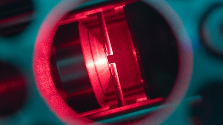
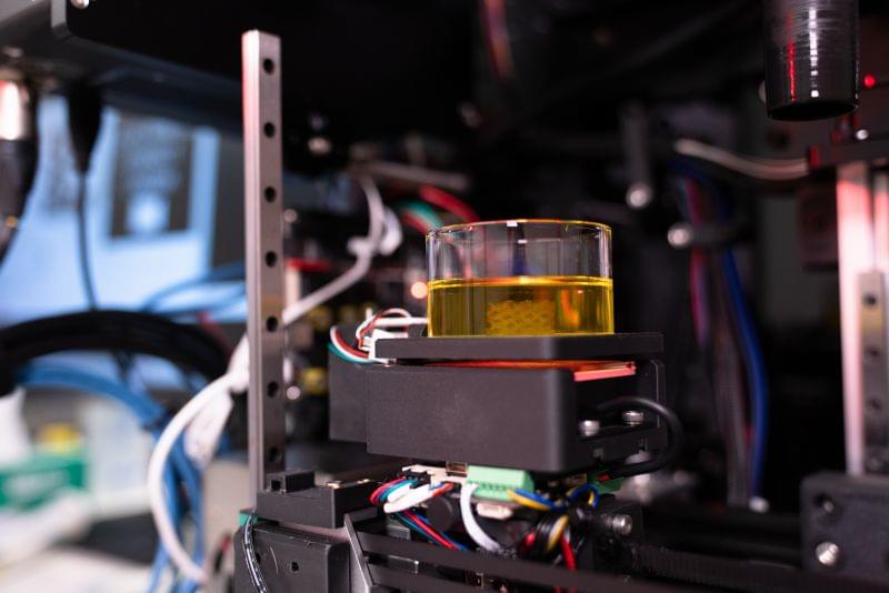
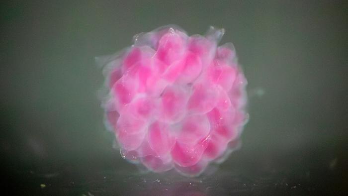
Volumetric 3D printing is an exciting technology that could lead to extremely rapid production of 3D printed parts by curing every particle of the object at once. Now, researchers from Utrecht University are applying the process to bioprinting and have 3D printed functioning liver units at centimeter scale in less than 20 seconds. The results were published in Advanced Materials.

Nov 23, 2022

3D Printing in Breast Reconstruction: From Bench to Bed

Posted by in categories: 3D printing, bioengineering, bioprinting, biotech/medical, computing

Circa 2021:3


Surgical management of breast cancer often results in the absence of the breast. However, existing breast reconstruction methods may not meet the need for a replacement tissue. Tissue engineering with the use of emerging materials offers the promise of generating appropriate replacements. Three-dimensional (3D) printing technology has seen a significantly increased interest and application in medically-related fields in the recent years. This has been especially true in complex medical situations particularly when abnormal or complicated anatomical surgical considerations or precise reconstructive procedures are contemplated. In addition, 3D bio-printing which combines cells with bio-material scaffolds offers an exciting technology with significant applications in the field of tissue engineering. The purpose of this manuscript was to review a number of studies in which 3D printing technology has been used in breast reconstructive surgical procedures, and future directions and applications of 3D bio-printing.

Breast cancer is the most common cancer diagnosed among US women and is second only to lung cancer as a cause of cancer death among women as of 2019. Because ~268,600 (almost six times than DCIS) new cases prove to be an invasive type of breast cancer (1), many women had to choose the removal of the breast, with immediate consideration for a replacement tissue. Although this was satisfactory in many patients, either saline or gel-filled breast implants (2) do carry real risks of complications such as infection, capsular contracture, implant dislocation, or deformities (3, 4). The option of autologous reconstruction can be more texturally natural aesthetically, but it requires a more complex procedure, significant time and expense, and possible muscle weakness or hernia formation at the tissue donor site (5). Tissue engineering intends to address these limitations by combining the 3D printing technology with synthetic or natural structural elements.

Continue reading “3D Printing in Breast Reconstruction: From Bench to Bed” »

Nov 23, 2022

Landmark Transplant Turns 3D Bioprinting on Its Ear

Posted by in categories: 3D printing, bioprinting, biotech/medical

A reconstructed ear made of living cells could herald a new era of 3D-printed organs.

Nov 13, 2022

Uganda’s New Satellite Contains Equipment to 3D Print Human Tissue in Orbit

Posted by in categories: 3D printing, bioprinting, biotech/medical, food, security

With the help of NASA and Japan, Uganda has officially become a spacefaring nation — and its newly-launched PearlAfricaSat-1 craft has some pretty nifty tech onboard.

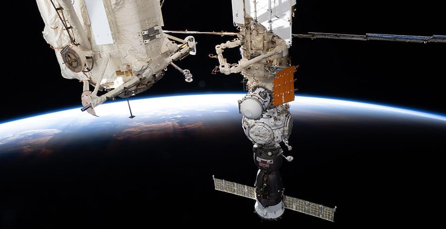
As the Uganda-based Nile Post reports, the satellite launched out of NASA’s Mid-Atlantic Regional Spaceport facility in Virginia on the morning of November 7 will not only provide important agricultural and security monitoring features for the developing nation, but will also conduct experiments involving the 3D printing of human tissue.

Per the Ugandan news site, the tissues printed on PearlAfricaSat-1 will be used in research into the effects microgravity has on ovary function — and as Quartz notes in its write-up of the NASA and Japan-supported mission, the microgravity aspect of the experiments is key because “bioprinting” human organs is difficult to achieve with Earth’s gravity.

Oct 14, 2022

3D printing plant cells shows promise for studying cell function

Posted by in categories: 3D printing, bioprinting

A new study from North Carolina State University shows a reproducible way of studying cellular communication among varied types of plant cells by “bioprinting” these cells via a 3D printer. Learning more about how plant cells communicate with each other—and with their environment—is key to understanding more about plant cell functions and could ultimately lead to creating better crop varieties and optimal growing environments.

The researchers bioprinted cells from the model plant Arabidopsis thaliana and from soybeans to study not just whether plant cells would live after being bioprinted—and for how long—but also to examine how they acquire and change their identity and function.

“A plant root has a lot of different cell types with specialized functions,” said Lisa Van den Broeck, an NC State postdoctoral researcher who is the first author of a paper describing the work. “There are also different sets of genes being expressed; some are cell-specific. We wanted to know what happens after you bioprint and place them into an environment that you design: Are they alive and doing what they should be doing?”

Jun 29, 2022

Using colloidal nanodiscs for 3D bioprinting tissues and tissue models

Posted by in categories: 3D printing, bioprinting, biotech/medical, engineering, nanotechnology

Extrusion-based 3D printing/bioprinting is a promising approach to generating patient-specific, tissue-engineered grafts. However, a major challenge in extrusion-based 3D printing and bioprinting is that most currently used materials lack the versatility to be used in a wide range of applications.

New nanotechnology has been developed by a team of researchers from Texas A&M University that leverages colloidal interactions of nanoparticles to print complex geometries that can mimic tissue and organ structure. The team, led by Dr. Akhilesh Gaharwar, associate professor and Presidential Impact Fellow in the Department of Biomedical Engineering, has introduced colloidal solutions of 2D nanosilicates as a platform technology to print complex structures.

2D nanosilicates are disc-shaped inorganic nanoparticles 20 to 50 nanometers in diameter and 1 to 2 nanometers in thickness. These nanosilicates form a “house-of-cards” structure above a certain concentration in water, known as a colloidal solution.

Jun 11, 2022

When we’ll be able to 3D-print organs and who will be able to afford them

Posted by in categories: 3D printing, bioprinting, biotech/medical

To begin the process of bioprinting an organ, doctors typically start with a patient’s own cells. They take a small needle biopsy of an organ or do a minimally invasive surgical procedure that removes a small piece of tissue, “less than half the size of a postage stamp,” Atala said. “By taking this small piece of tissue, we are able to tease cells apart (and) we grow and expand the cells outside the body.”

This growth happens inside a sterile incubator or bioreactor, a pressurized stainless steel vessel that helps the cells stay fed with nutrients – called “media” – the doctors feed them every 24 hours, since cells have their own metabolism, Lewis said. Each cell type has a different media, and the incubator or bioreactor acts as an oven-like device mimicking the internal temperature and oxygenation of the human body, Atala said.

“Then we mix it with this gel, which is like a glue,” Atala said. “Every organ in your body has the cells and the glue that holds it together. Basically, that’s also called ‘extracellular matrix.’”.

Page 4 of 1712345678Last