Toggle light / dark theme

Sound-sensing hair bundles in our ears act as tiny thermodynamic machines

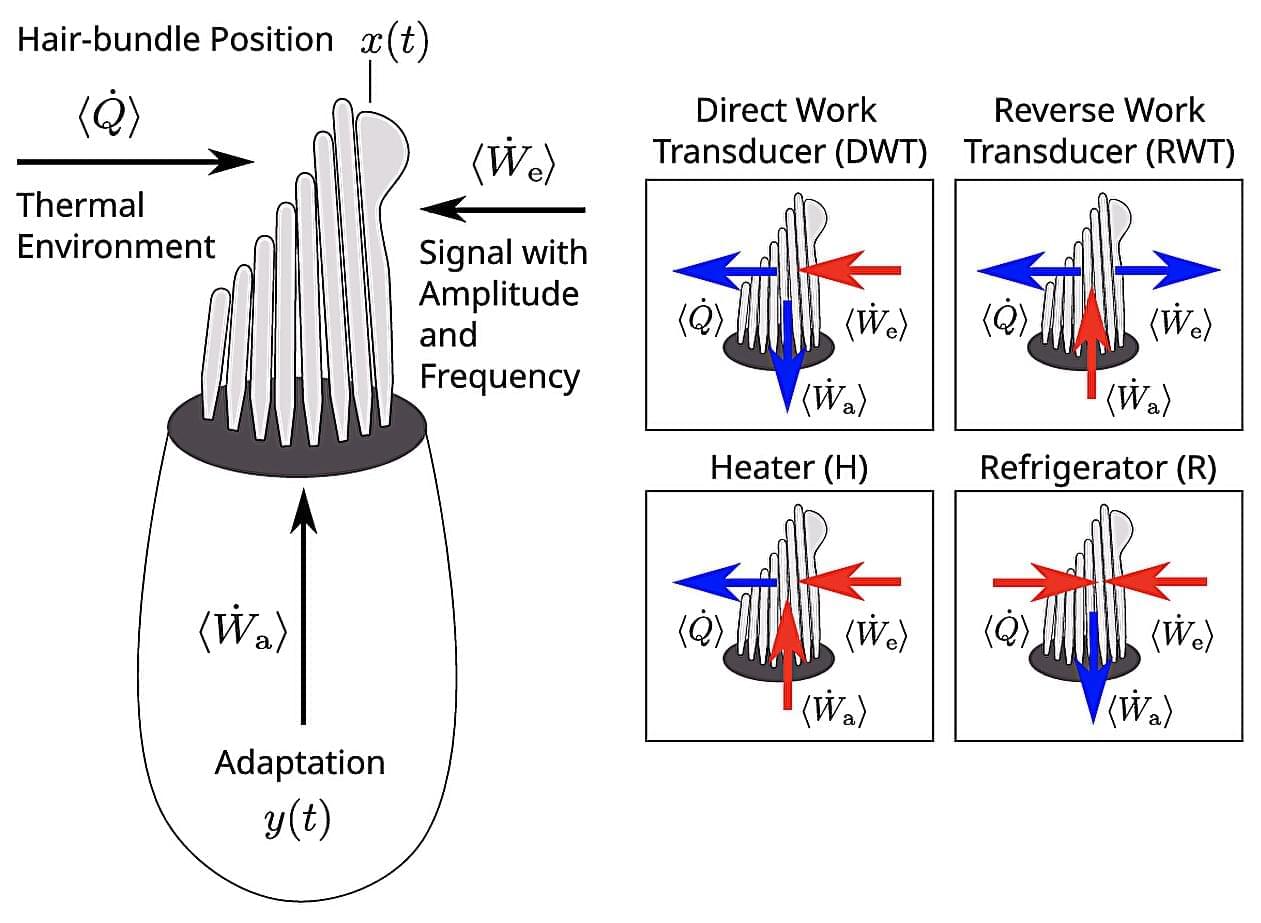
The hair cells lining the inner ear are among the most sophisticated structures in the human body: capable of detecting sounds as faint as a whisper, while helping to maintain our sense of balance. Through new models detailed in PRX Life, a team led by Roman Belousov at the European Molecular Biology Laboratory has revealed for the first time how oscillating bundles attached to these cells operate in different thermodynamic regimes—offering a new framework for understanding how our hearing works at a fundamental level.

Within the inner ear, each hair cell hosts a hair “bundle”: a cluster of tiny, bristle-like projections that vibrate in response to incoming sound waves. The mechanical energy from these oscillations is then converted into electrical signals which travel to the brain. Rather than being passive receivers, these bundles actively oscillate —driven by molecular motors within the cell that allow them to amplify faint signals and tune in to specific frequencies.

But despite decades of study, researchers are still unclear on the connection between this active oscillation and the hair bundle’s response to external sound. Existing models tended to treat bundles as if they were moving spontaneously, without accounting for what happens when they actually interact with sound.

AI uncovers hidden immune defenses inside bacteria

Researchers at the Massachusetts Institute of Technology (MIT) have discovered thousands of new proteins that protect bacteria from virus attacks using an AI system called DefensePredictor. What would usually take months of lab work can now be narrowed down to promising candidates in minutes.

Bacteria are under constant attack from viruses called bacteriophages. One of their most powerful defenses is CRISPR-Cas, a system that cuts up viral DNA to stop an infection and is now a valuable biotechnology tool for precisely editing genes in a lab.

Traditional methods of finding these defenses are long and laborious, equivalent to looking for a needle in a haystack. They involve searching for nearby known defensive genes and manually testing thousands of DNA fragments. But now, AI can take the strain.

Key protein found to protect cartilage, offering new hope for osteoarthritis treatment

Osteoarthritis, a condition that causes pain and reduced mobility in joints such as the knees and fingers, is one of the most common joint disorders worldwide, particularly among aging populations. The disease is characterized by the gradual breakdown of cartilage, which normally cushions the bones within joints.

Despite its prevalence, current treatments for osteoarthritis mainly focus on alleviating pain rather than addressing the underlying cause of cartilage degeneration. Effective therapies that can halt or reverse cartilage damage remain limited.

A joint research team led by Dr. Chul-Ho Lee and Dr. Yong-Hoon Kim at the Laboratory Animal Resource Center of the Korea Research Institute of Bioscience and Biotechnology (KRIBB), in collaboration with Prof. JinHyun Kim at Chungnam National University Hospital, has identified a key protein, SHP (NR0B2), that plays a critical protective role in cartilage and may offer a new therapeutic strategy for osteoarthritis. The paper is published in the journal Nature Communications.

DNA polymerase μ protects macrophages from DNA damage produced during pro-inflammatory activation

Polμ is induced in macrophages by ROS upon pro-inflammatory stimuli and is essential for efficient repair of DNA double-strand breaks. Its deficiency compromises macrophage survival and persistence in inflammatory models of infection and tissue repair, underscoring Polμ’s critical role in counteracting ROS-mediated genotoxic stress and in achieving a correct inflammatory resolution.

Metabolic dysfunction-associated steatotic liver disease and steatohepatitis-associated hepatocarcinoma preclinical models

Preclinical models are essential to study disease pathogenesis and test novel treatments. Here, a broad overview ofis provided, detailing main features, advances and limitations of in vitro and in vivo models, and how they translate to human disease.

Mapping mutations at scale in a single gene reveals new neurodevelopmental condition

The ability of different genetic variants—changes to one or more building blocks of DNA—to cause disease, and to what extent, has historically been opaque. Geneticist and Crick group leader Greg Findlay has pioneered a new method in the hope of changing this. Called “saturation genome editing,” the new technique involves mapping every single variant in a given gene to work out what it does and pinpoint which changes are responsible for specific disorders.

While Greg was refining these experiments, Nicky Whiffin, associate professor at the University of Oxford, had identified that mutations in a tiny gene were behind a rare inherited neurodevelopmental disorder, known as ReNU syndrome, which impacts brain function, development and motor skills. Children develop this syndrome if a single copy of the RNU4-2 gene is mutated in a specific way.

Nicky initially found that several distinct mutations in a critical region of the gene caused the condition, and she was keen to understand if some of these genetic variants led to more severe disease.

Mapping pesticide mixtures to cancer risk at the country scale with spatial exposomics

Using an integrative spatial Bayesian framework that merges high-resolution environmental pesticide risk modelling with comprehensive cancer registry data, this analysis reveals spatial patterns of pesticide exposure and liver tissue-derived molecular signatures across Peru, establishing links between pesticide usage and cancer insurgence at the national scale.

ADAMTS-7 Vaccine Confers Renal and Vascular Protection in Chronic Kidney Disease

Yang & colleagues demonstrate a peptide vaccine targeting the metalloproteinase ADAMTS7 mitigates the damage to the kidneys & blood vessels related to chronic kidney disease. Learn more at.


ELetters should relate to an article recently published in the journal and are not a forum for providing unpublished data. Comments are reviewed for appropriate use of tone and language. Comments are not peer-reviewed. Acceptable comments are posted to the journal website only. Comments are not published in an issue and are not indexed in PubMed. Comments should be no longer than 500 words and will only be posted online. References are limited to 10. Authors of the article cited in the comment will be invited to reply, as appropriate.

Abstract: Do cells of different lineages age differently?

https://doi.org/10.1172/JCI195772 Sundeep Khosla & team compare the senescence phenotype of mesenchymal versus immune cells from murine bone and bone marrow, revealing important differences between them.

The figure shows mesenchymal cells exhibit higher absolute levels of senescence signatures than immune cells.


Address correspondence to: Madison L. Doolittle, Center for Regenerative Medicine and Skeletal Development, UConn Health, 263 Farmington Avenue, Farmington, Connecticut 6,030, USA. Phone: 860.679.1757; Email: [email protected]. Or to: Sundeep Khosla, Guggenheim 7, Mayo Clinic College of Medicine, 200 First Street SW, Rochester, Minnesota 55,905, USA. Phone: 507.255.6663; Email: [email protected].

Histone modification clocks for robust cross-species biological age prediction and elucidating senescence regulation

Building upon these insights, we constructed 36 histone modification-based epigenetic clocks, which exhibited robust predictive accuracy (mean Pearson’s r = 0.91) across multiple tissues and marks. Among these, the blood-derived H3K27ac clock emerged as a particularly powerful model, outperforming several established DNA methylation clocks under matched conditions. This performance is remarkable considering that DNA methylation clocks have undergone extensive optimization over the past decade (9, 16, 18), while our histone-based approach represents a first-generation effort.

A distinctive advantage of our histone-based clocks is their resilience to technical and biological noise. When exposed to artificial Gaussian noise, the histone-based clock maintained stable predictive performance, in contrast to the sharp degradation observed in many methylation-based models. This robustness is likely attributable to the broader, structural nature of histone mark signals, which may be less sensitive to local fluctuations than single CpG methylation values. This characteristic makes histone clocks potentially more suitable for noisy, heterogeneous, or clinically derived datasets where sample quality may vary.

The practical utility of our histone-based clocks was further demonstrated by their ability to detect biological age acceleration in leukemia samples and capture age reversal following therapeutic interventions. These applications highlight the potential of histone-based clocks as biomarkers for disease states and treatment responses, offering a complementary approach to existing clinical tools.

/* */