Toggle light / dark theme

Arrays of quantum rods could enhance TVs or virtual reality devices, research suggests

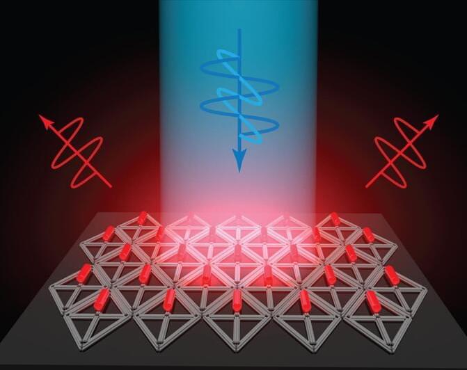
Flat screen TVs that incorporate quantum dots are now commercially available, but it has been more difficult to create arrays of their elongated cousins, quantum rods, for commercial devices. Quantum rods can control both the polarization and color of light, to generate 3D images for virtual reality devices.

Using scaffolds made of folded DNA, MIT engineers have come up with a new way to precisely assemble arrays of quantum rods. By depositing quantum rods onto a DNA scaffold in a highly controlled way, the researchers can regulate their orientation, which is a key factor in determining the polarization of light emitted by the array. This makes it easier to add depth and dimensionality to a virtual scene.

“One of the challenges with quantum rods is: How do you align them all at the nanoscale so they’re all pointing in the same direction?” says Mark Bathe, an MIT professor of biological engineering and the senior author of the new study. “When they’re all pointing in the same direction on a 2D surface, then they all have the same properties of how they interact with light and control its polarization.”

New method a step toward future 3D printing of human tissues

A team of bioengineers and biomedical scientists from the University of Sydney and the Children’s Medical Research Institute (CMRI) at Westmead have used 3D photolithographic printing to create a complex environment for assembling tissue that mimics the architecture of an organ.

The teams were led by Professor Hala Zreiqat and Dr. Peter Newman at the University of Sydney’s School of Biomedical Engineering and developmental biologist Professor Patrick Tam who leads the CMRI’s Embryology Research Unit. Their paper was published in Advanced Science.

Using bioengineering and cell culture methods, the technique was used to instruct stem cells derived from or to become specialized cells that can assemble into an organ-like structure.

Japanese Scientists Construct Complex 3D Organoids With Ingenious Device

A team of scientists led by Masaya Hagiwara of RIKEN national science institute in Japan has developed an ingenious device, using layers of hydrogels in a cube-like structure, that allows researchers to construct complex 3D organoids without using elaborate techniques. The group also recently demonstrated the ability to use the device to build organoids that faithfully reproduce the asymmetric genetic expression that characterizes the actual development of organisms. The device has the potential to revolutionize the way we test drugs, and could also provide insights into how tissues develop and lead to better techniques for growing artificial organs.

Scientists have long struggled to create organoids—organ-like tissues grown in the laboratory—to replicate actual biological development. Creating organoids that function similarly to real tissues is vital for developing medicines since it is necessary to understand how drugs move through various tissues. Organoids also help us gain insights into the process of development itself and are a stepping stone on the way to growing whole organs that can help patients.

A Drug For Regrowing Teeth Could Be Available Within The Next Decade

Teeth don’t grow back once we become adults: any wear and tear is permanent – as those of us with fillings will know – which is why it’s important to keep them as clean and healthy as we can.

However, this is something scientists are now looking to change.

It’s been announced that clinical trials for a potential tooth regrowth treatment are set to begin in July 2024, building on decades of research in the field. If those trials are successful, therapeutic drugs could be available by 2030.

MaxCyte signs strategic platform license with prime medicine to advance next-generation gene editing therapies for patients

MaxCyte, Inc., a leading, cell-engineering focused company providing enabling platform technologies to advance the discovery, development and commercialization of next-generation cell-based therapeutics and to support innovative, cell-based research, today announced the signing of a strategic partnership with Prime Medicine, Inc., a biotechnology company committed to delivering a new class of differentiated one-time curative genetic therapies.

New Biomarkers Improve Diagnostics for Multiple Sclerosis & An MS-Like Disorder

Multiple sclerosis (MS) can cause a huge range of symptoms in different patients, and the severity can vary dramatically. It is an inflammatory condition in which the body attacks myelin sheaths, a protective insulation surrounding nerve cells. This can cause fatigue, pain, paralysis, and symptoms that gradually get worse. MS can be very difficult to diagnose because the symptoms can be so different in different patients, and the presence of brain lesions is the clearest indication of the disease. MRIs that can reveal those brain lesions are only useful once the disease have progressed to the point of brain damage, however.

The innate immune system presents a potential option for monitoring the progression of MS. The disease causes inflammation, so researchers tracked immune cells in the brain called macrophages, and assessed brain inflammation in a mouse model of MS. The findings have been reported in Science Translational Medicine.

/* */