“Pets have more love and compassion in them than most humans.”–Robert Wagner.
The COVID-19 pandemic didn’t just transform how we work and communicate. It also accelerated the need for more proactive health measures for chronic health problems tied to diet. Such problems have emerged as a top risk factor for coronavirus and people with poor metabolic health accounted for half of COVID-19 hospitalizations in some regions around the world. The resulting high numbers led the authors of a report in The Lancet to issue a call for more resources to tackle metabolic health to avoid needless deaths.
Thankfully, new tools have been developed to offer comprehensive understanding of nutrition. This expertise and technology won’t just help us tackle metabolic health – it could help us finally fully realize the power of plants to improve health and wellness outcomes.

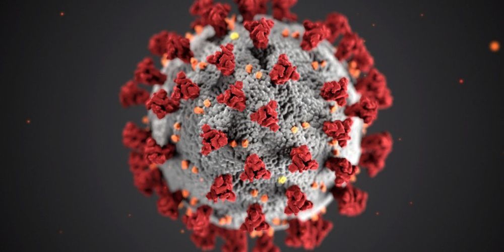
Coronavirus disease (COVID-19) is an emerging global pandemic that is threatening the viability of healthcare systems worldwide. The virus responsible for the disease is also known as severe acute respiratory syndrome coronavirus 2 and abbreviated as SARS-CoV-2. The eruption started in China on December 29, 2019 and by March 2020 it was reported that it had spread to several countries across the world.

In the final days of 2019, Chinese doctors identified a number of similar cases of pneumonia in the city of Wuhan in China. Wuhan is the capital city of Hubei Province in China accommodating 11 million inhabitants. Soon, it was discovered that the disease was caused by a new strain of virus which was named SARS-CoV-2.

Looks like the Centers for Disease Control and Prevention (CDC) has found the root cause of the latest Salmonella Newport outbreak. And it’s not alien DNA or demon sperm. It’s onions, specifically red onions.
Yep, if you like red onions with your salads, on your pasta, in your burgers, or just all over your body, you may be shedding a tear. Eating red onions is the one thing that many people affected by this Salmonella outbreak seem to have in common. Well, that and diarrhea as well as all the other wonderful stuff that comes with Salmonella infections.
I first covered this outbreak eight days ago for Forbes. Back then, the cause of the outbreak, which had already affected at least 125 people in 15 states at the time, was unknown. The CDC couldn’t warn the public to avoid any specific foods, and avoiding all foods would not have been a practical suggestion.
Do you know what natural ionising radiation is? Where can you find natural resources of radiation? What are the levels of natural sources of radiation in Europe? Do you know the pathways of ionising radiation? Natural radionuclides, both terrestrial and cosmogenic, migrate in the environment through different pathways: air, water, rock, soil and the food chain. Radionuclides may then enter the.
Human body through ingestion (food and drinking water) and inhalation giving, so-called, internal exposure. External exposure is due to cosmic radiation and radiation from terrestrial radionuclides present in soil, rock and building materials. The first ever ‘European atlas of natural radiation’ uses informative texts, stunning photographs and striking maps to answer and explain these and other questions.
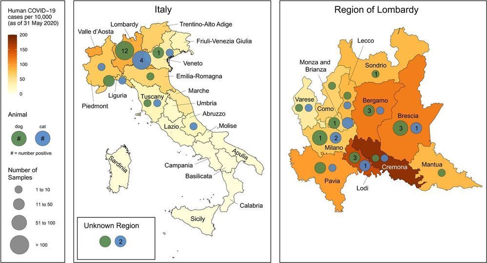
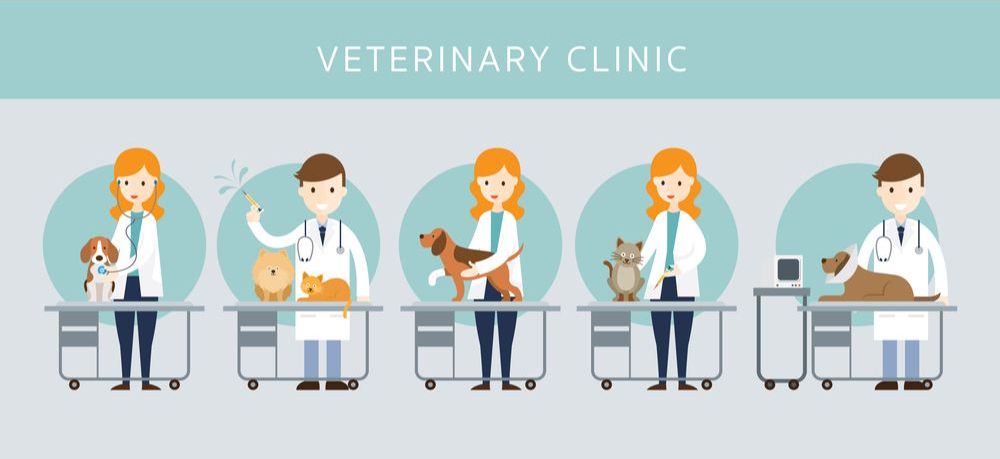
SARS-CoV-2 originated in animals and is now easily transmitted between people. Sporadic detection of natural cases in animals alongside successful experimental infections of pets, such as cats, ferrets and dogs, raises questions about the susceptibility of animals under natural conditions of pet ownership. Here we report a large-scale study to assess SARS-CoV-2 infection in 817 companion animals living in northern Italy, sampled at a time of frequent human infection. No animals tested PCR positive. However, 3.4% of dogs and 3.9% of cats had measurable SARS-CoV-2 neutralizing antibody titers, with dogs from COVID-19 positive households being significantly more likely to test positive than those from COVID-19 negative households. Understanding risk factors associated with this and their potential to infect other species requires urgent investigation.
One Sentence Summary SARS-CoV-2 antibodies in pets from Italy.
Severe acute respiratory syndrome coronavirus 2 (SARS-CoV-2) emerged in late December 2019 in Wuhan, Hubei province, China , possibly as a spillover from bats to humans , and rapidly spread worldwide becoming a pandemic. Although the virus is believed to spread almost exclusively by human-to-human transmission, there are concerns that some animal species may contribute to the ongoing SARS-CoV-2 pandemic epidemiology. To date, sporadic cases of SARS-CoV-2 infection have been reported in dogs and cats. These include detection of SARS-CoV-2 RNA in respiratory and/or fecal specimens of dogs and cats with or without clinical signs (5−7), as well as of specific antibodies in sera from pets from coronavirus disease 2019 (COVID-19) affected areas (7,8).
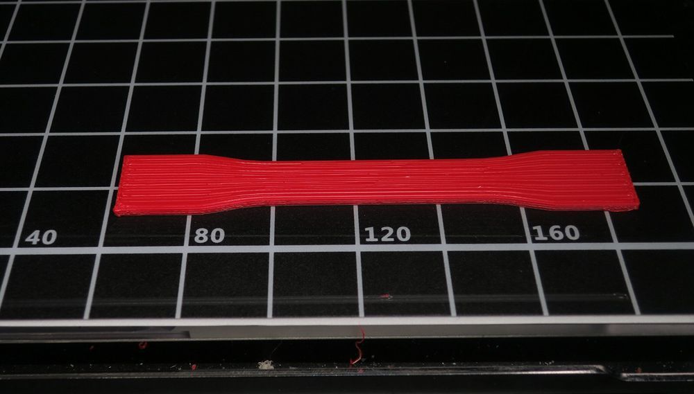
More durable prosthetics and medical devices for patients and stronger parts for airplanes and automobiles are just some of the products that could be created through a new 3D printing technology invented by a UMass Lowell researcher.
Substances such as plastics, metals and wax are used in 3D printers to make products and parts for larger items, as the practice has disrupted the prototyping and manufacturing fields. Products created through the 3D printing of plastics include everything from toys to drones. While the global market for 3D plastics printers is estimated at $4 billion and growing, challenges remain in ensuring the printers create objects that are produced quickly, retain their strength and accurately reflect the shape desired, according to UMass Lowell’s David Kazmer, a plastics engineering professor who led the research project.
Called injection printing, the technology Kazmer pioneered is featured in the academic journal Additive Manufacturing posted online last week.

The Stanford team worked with researchers at the Department of Energy’s Lawrence Berkeley National Laboratory to develop a technique called prophylactic antiviral CRISPR in human cells, or PAC-MAN. The technology disables viruses by scrambling their genetic code. The researchers developed a new way to deliver the technology into lung cells, they reported in the journal Cell.
Stanford bioengineers teamed up with researchers at the Lawrence Berkeley National Laboratory to develop a CRISPR system that neutralizes SARS-CoV-2 by scrambling the virus’s genetic code. They believe the technology could prove useful for combating several types of viruses, including influenza.