Lyme disease can be easiest to treat in its earliest stages, but current tests often miss infections during that critical window and cannot tell whether bacteria are still present or were cleared years ago. New research led by Tufts University School of Medicine suggests that a group of immune molecules called anti-lipid antibodies may address these shortcomings.
The findings, published in Infection and Immunity, could lead to improved tests that identify Lyme disease earlier, when antibiotics can best prevent more debilitating disease. They also may help clinicians better identify patients who continue to experience symptoms of infection after treatment—and potentially find new drug targets to help them.
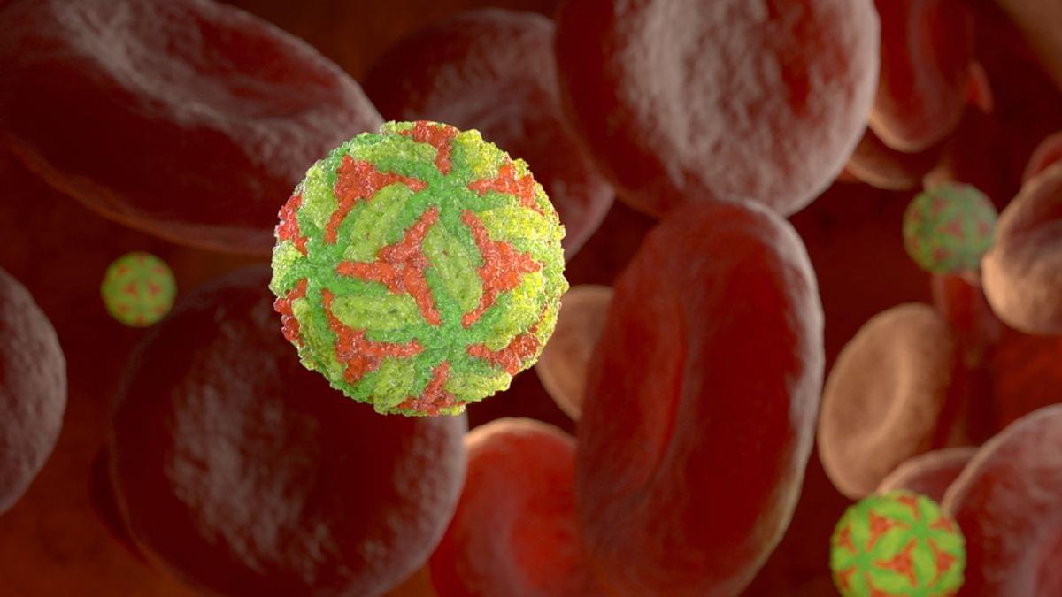
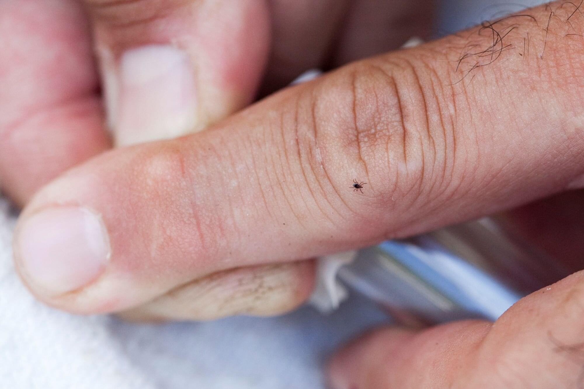
Nearly half a million Americans are diagnosed and treated for Lyme disease each year. Caused by the bacterium Borrelia burgdorferi and spread through the bite of infected blacklegged ticks (also known as deer ticks), the disease can lead to arthritis, neurological problems and heart complications if untreated.