In healthy aging strategies, nutritional supplements synergize with optimized dietary and lifestyle interventions by modulating aging-related molecular pathways.[ 8, 9 ] Notably, NMN exerts multi-organ anti-aging effects by elevating NAD+ levels to activate the SIRT1 pathway, thereby significantly enhancing mitochondrial function while reducing oxidative stress and DNA damage.[ 10 ] Similarly, curcumin delays aging and related diseases through pleiotropic mechanisms involving oxidative stress regulation, anti-inflammatory actions, telomere maintenance, and sirtuin protein modulation.[ 11 ] However, practical applications face significant challenges: bioactive compounds like resveratrol and curcumin suffer from limited bioavailability due to poor aqueous solubility and first-pass metabolism, while excessive supplementation of antioxidants such as vitamins C/E may disrupt reactive oxygen species (ROS) signaling homeostasis, potentially inducing cellular toxicity or even increasing hemorrhagic risk.[ 12-14 ] Future development of anti-aging supplements should focus on: 1) innovative formulation strategies to enhance bioavailability; 2) optimized dosing regimens to minimize toxicity; and 3) long-term clinical studies to validate efficacy.
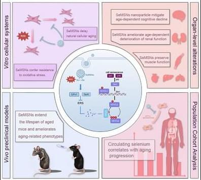
Selenium, an essential trace element with diverse biological activities, plays a critical role in healthy aging.[ 15-17 ] ≈1 billion people worldwide are affected by selenium deficiency, which is closely linked to neurological disorders, cardiovascular abnormalities, malignancies, and immune dysfunction.[ 18-20 ] Substantial evidence supports the anti-aging effects of selenium through multiple mechanisms: 1) Selenomethionine (SeMet) effectively suppresses Fe2+/H2O2- or Aβ-induced free radical generation, demonstrating therapeutic potential for Alzheimer’s disease characterized by oxidative stress;[ 21 ] 2) Selenium supplementation elevates serum GPx3 levels, a selenoprotein predominantly localized in the basement membrane of renal proximal tubules, modulating mitochondrial quality control pathways to mitigate heavy metal-induced renal aging;[ 22 ] and 3) Our recent findings reveal that selenium supplementation significantly attenuates age-related muscle atrophy by preserving redox homeostasis and regulating muscle protein metabolism.[ 23 ] Recent clinical trials in patients with advanced non-small cell lung cancer (NSCLC) demonstrated that oral administration of selenium nanoparticles (SeNPs) as a dietary supplement (200 µg day−1) in combination with Bev+AP chemotherapy significantly enhanced therapeutic outcomes compared to chemotherapy alone. The SeNPs combination group showed remarkable tumor regression, with progression disease rates decreasing dramatically from 50% to 0% and partial response rates increasing to 83.3%, along with significantly improved objective response rate and disease control rate.[ 24 ] Importantly, this regimen maintained excellent safety profiles without triggering fluctuations in pro-inflammatory or immunosuppressive cytokines. These compelling findings not only establish SeNPs as a safe and effective adjuvant therapy for advanced NSCLC but also provide valuable clinical translation data for nano-selenium formulations in oncology. Despite selenium’s proven benefits in reducing oxidative damage, maintaining genomic stability, and delaying telomere shortening, its narrow therapeutic window, limited bioavailability, and specific mechanisms in multi-organ protection during natural aging require further investigation.
Nanodelivery carriers have emerged as a next-generation platform for gene and drug delivery, offering tunable physicochemical properties such as size, composition, and surface modifications.[ 25 ] Our team has developed organically-bridged mesoporous silica nanoparticles (MSNs) by incorporating functional diselenide bonds into the silica framework at the molecular level, addressing the critical challenge of poor biodegradability in conventional silica materials.[ 26 ] This nanocarrier exhibits unique dual redox-responsive properties, allowing for more precise maintenance of redox homeostasis compared to traditional antioxidants, aligning with the core goal of preserving organismal homeostasis in anti-aging research. Building on this breakthrough, a comprehensive research framework was established: first, this study constructed a natural aging mouse model with MSNs, disulfide-bridged MSNs (SMSNs), commercially available SeMet as controls and then compared the effects of diselenide-bridged MSNs (SeMSNs) on lifespan extension, frailty delay, and multi-organ anti-aging. Next, key pathways and targets were identified through multi-organ transcriptome sequencing, followed by in-depth mechanistic studies. Finally, clinical translation was integrated by analyzing the correlation between serum selenium levels and aging biomarkers in the elderly, and validating the clinical effects of SeMSNs using primary adipose precursor cells (APCs) models. This systematic approach provides a solid theoretical foundation and clinical evidence for the application of nano-selenium in anti-aging research.