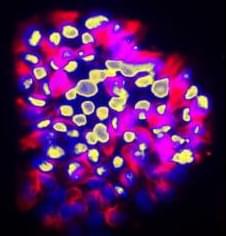
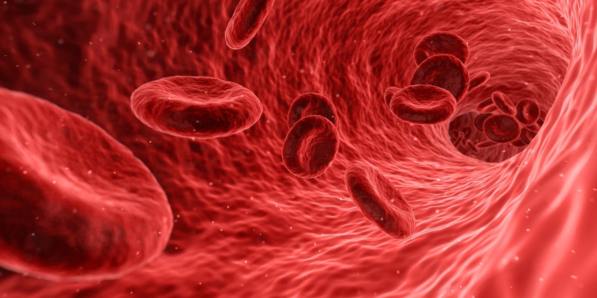
Toggle light / dark theme

Structures of hantavirus glycoprotein tetramers

This surface protein complex for the Andes virus is a mushroom-shaped structure called a Gn-Gc tetramer. To map the 3D structures, the team first produced virus-like particles that mimic a real virus, but without the genome that makes a virus infectious. They then used a cryo-electron microscope—which shines an electron beam through a frozen sample and detects the shadows created by molecules—to reconstruct the three-dimensional structures of the Gn-Gc tetramers on the surface of the virus-like particles.

But there was a twist: To obtain extremely high-resolution structures, the researchers painstakingly identified and isolated shadows from only the tetramers that were pointing sidewise relative to the electron beam and ignored those pointing in other directions. This allowed them to borrow a reconstruction method typically used on individual proteins.

The resulting structures have an extremely high resolution of 2.3 angstroms, meaning details the size of just a couple of atoms were effectively captured. That’s enough to represent a transformational improvement over another team’s model of the tetramer from a few years ago, at a resolution of 12 angstroms, still tiny but large enough to produce some key inaccuracies – ones effectively corrected with the newer method and resulting structure.

These latest structures show the Gn-Gc tetramer in a particular state before it has infected a cell. For vaccines or antibody therapies to be most effective against a hantavirus, mimicking surface proteins at this pre-infection stage is essential. ScienceMission sciencenewshighlights.


Hantaviruses, transmitted from rodents to people, have a death rate approaching 40%. They’re found around the world, and because there are no approved vaccines or treatments, they’re among the pathogens of highest concern for future pandemics. They made news in the United States last year when Betsy Arakawa, the wife of actor Gene Hackman, died from a hantavirus infection in New Mexico in March.

New findings published in the journal Cell about the Andes virus, a hantavirus endemic to the southwestern U.S. and other parts of North and South America, represent a crucial first step towards much-needed vaccines and antibody therapies for this and other hantaviruses.

A new class of Alzheimer’s biomarkers: Why protein shape may beat protein levels

Researchers have identified a new type of blood-based biomarker test for Alzheimer’s disease that measures structural changes in proteins, providing more information on the underlying biology of the disease than standard blood tests. The findings, published in Nature Aging, also provide new insights into how Alzheimer’s disease biology may differ between males and females.

“This work introduces a fundamentally new, blood-based approach to detecting and staging Alzheimer’s disease,” said Dr. Richard Hodes, director of NIH’s National Institute on Aging (NIA). “By revealing protein structural changes associated with genetic risk, symptom severity, and sex differences—features not captured by existing biomarkers—this research could enable earlier diagnosis and more effective clinical trials.”

A biotech company just doubled the lifespan of mice without changing their diet and without editing their genes

Instead, they trained the immune system to hunt down and destroy the cells that make the body age. Then they flooded the body with fresh stem cells to rebuild what was lost.

This isn’t science fiction. It’s longevity science happening right now.

Read more

Genomic reorganization at the transition to gametogenesis

Using a technique called Hi-C analysis, which looks at how DNA is arranged in three dimensions inside the nucleus, the team found that at this transitional point the genome’s three-dimensional organisation becomes less structured and chromosomes become more separated inside the nucleus.

Creating sperm and eggs in the laboratory (in vitro) remains one of the greatest challenges in reproductive biology. To study this process, scientists use primordial germ cell–like cells (PGCLCs), which are lab-generated cells derived from embryonic stem cells that mimic the embryo’s earliest reproductive cells. However, these PCGLCs often fail to complete all the steps of meiosis, making it difficult to create functional sperm and eggs in petri dishes.

After studying the process in germ cells from the embryos, the team studied lab-generated mouse PCGLCs to see if the centromeres migrated to the periphery of the nucleus in vitro too, but they did not see the same phenomenon.

“The presence of this chromosome conformation in embryonic germ cells, but not lab-grown cells, suggests that this structural change could be required for meiosis to proceed properly, and could explain why meiosis is so difficult to recreate outside the body,” says the author, “but we need to do more work to fully characterise the process before we can say for sure.”

“Our study has uncovered a previously unknown and frankly very surprising restructuring of genome architecture that occurs in developing germ cells, which we believe is critical for a successful execution of meiosis,” says the senior author. ScienceMission sciencenewshighlights.


In our cells, our DNA carries chemical or ‘epigenetic’ marks that decide how genes will be used in different tissues. Yet in the group of specialised cells, known as ‘germ cells’, which will later form sperm and eggs, these inherited chemical instructions must be erased or reshuffled so development can begin again with a fresh blueprint in future generations.

Foundation AI model uses MRI data to predict multiple brain disorders

Artificial intelligence (AI) systems are computational models that can learn to identify patterns in data, make accurate predictions or generate content (e.g., texts, images, videos or sound recordings). These models can reliably complete various tasks and are now also used to carry out research rooted in different fields.

Over the past few decades, some AI models have proved promising for the early diagnosis and study of specific diseases or neuropsychiatric conditions. For instance, by analyzing large amounts of brain scans collected using a noninvasive technique known as magnetic resonance imaging (MRI), AI could uncover patterns associated with tumors, strokes and neurodegenerative diseases, which could help to diagnose these conditions.

Researchers at Mass General Brigham, Harvard Medical School and other institutes recently developed Brain Imaging Adaptive Core (BrainIAC), a large AI system pre-trained on a vast pool of MRI data that could be adapted to tackle different tasks. This foundation model, presented in a paper published in Nature Neuroscience, was found to outperform many models that were trained to complete specific medical or neuroscience-related tasks.

Protein Folding and Quality Control in the Endoplasmic Reticulum: Recent Lessons from Yeast and Mammalian Cell Systems

The evolution of eukaryotes was accompanied by an increased need for intracellular communication and cellular specialization. Thus, a more complex collection of secreted and membrane proteins had to be synthesized, modified, and folded. The endoplasmic reticulum (ER) thereby became equipped with devoted enzymes and associated factors that both catalyze the production of secreted proteins and remove damaged proteins. A means to modify ER function to accommodate and destroy misfolded proteins also evolved. Not surprisingly, a growing number of human diseases are linked to various facets of ER function. Each of these topics will be discussed in this article, with an emphasis on recent reports in the literature that employed diverse models.

Adaptive Optimization of Vascular-Targeted Photodynamic Therapy Efficiency Based on Hyperspectral-Photoacoustic Dual-Modality Imaging Feedback

Objective: To enhance vascular-targeted photodynamic therapy (V-PDT) efficacy by integrating real-time dosimetric monitoring and adaptive irradiance modulation based on dynamic physiological feedback. Impact Statement: This study presents a closed-loop, dual-modality optical imaging-guided V-PDT platform that enables individualized, oxygen-informed irradiance control, improving therapeutic precision and efficiency. Introduction: While V-PDT is a promising, minimally invasive treatment for tumors and vascular abnormalities, its efficacy is often hindered by rapid oxygen depletion under high irradiance, leading to treatment-limiting hypoxia. Accurate, real-time assessment of both photosensitizer concentration and blood oxygenation is essential to guide optimized therapeutic strategies, yet such capability has remained elusive in clinical settings.

Abstract: In the 1960’s, megamitochondria in hepatocytes were identified in injured liver tissue

Here, Wen-Xing Ding find alterations in mitochondrial dynamics and the accumulation of large mitochondria contribute to liver tumor development in mice: https://doi.org/10.1172/JCI194441 # MASH

The EM image shows liver cells with megamitochondria (arrows) from mice lacking liver-specific dynamin-related protein 1 (Dnm1).


1Department of Pharmacology, Toxicology and Therapeutics, University of Kansas Medical Center, Kansas City, Kansas, USA.

2Department of Pharmaceutical Sciences, Eugene Applebaum College of Pharmacy and Health Sciences and.

3Department of Pharmacology, School of Medicine, Wayne State University, Detroit, Michigan, USA.

Paracrine Regulation and Immune System Pathways in the Inflammatory Tumor Microenvironment of Lung Cancer: Insights into Oncogenesis and Immunotherapeutic Strategies

Simple SummaryDespite massive strides taken across the board in oncology, there remain gaps in understanding the relationship between cancer cells and the body’s immune system, tissues, and signaling pathways.

/* */