Measles is a highly contagious viral infection that causes high fever, cough, red/watery eyes, and a characteristic blotchy rash, spreading through airborne droplets. It primarily affects children but can strike anyone, with severe cases leading to pneumonia, brain swelling, or death. Prevention is primarily through the MMR vaccine, which is 97% effective.
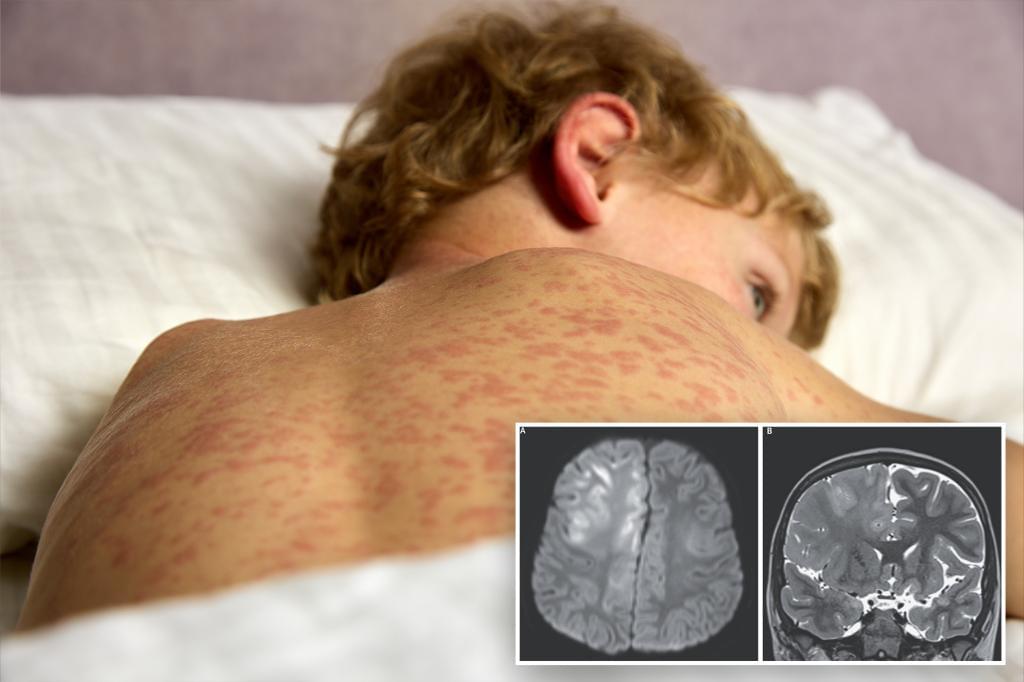
//He had contracted measles as a baby of just 7 months old — but fast-forward years later to when he was 6 and experiencing cognitive deterioration and seizures.
Doctors eventually diagnosed him with subacute sclerosing panencephalitis (SSPE), a neurological disease that can develop years after a measles infection.
This brain disorder usually starts with subtle personality changes, like memory loss, irritability or mood swings. Over time, it can progress to involuntary muscle spasms, loss of coordination, severe brain damage, coma — and almost always death.\
.
What is subacute sclerosing panencephalitis (SSPE): Subacute sclerosing panencephalitis (SSPE) is a rare, fatal, progressive neurodegenerative disorder of the central nervous system caused by a persistent, mutated measles virus infection. Typically affecting children or adolescents years after an initial infection, it causes cognitive decline, myoclonic jerks, and seizures, leading to death within 1–3 years. There is no cure, though prevention via measles vaccination is highly effective.
Even those who make a full recovery from the initial infection face a lurking threat: a deadly disease that remains latent until striking — and killing — years later.


