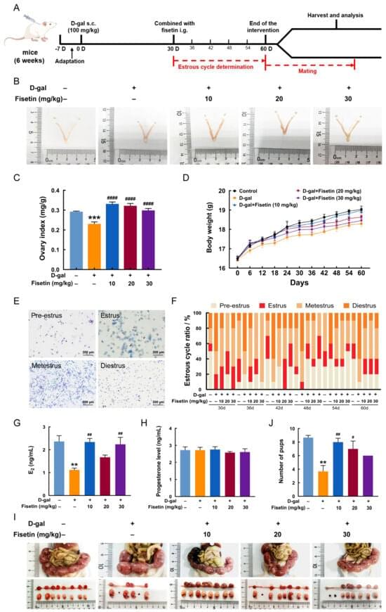
Toggle light / dark theme

Ferroptosis as a therapeutic target in glioblastoma: Mechanisms and emerging strategies

Ferroptosis: a promising therapeutic strategy in glioblastoma👇

✅Glioblastoma multiforme (GBM) is an aggressive brain tumor characterized by rapid growth and resistance to conventional therapies. Recent research highlights ferroptosis, a regulated form of cell death driven by iron-dependent lipid peroxidation, as a novel and promising approach for GBM treatment.

✅One key mechanism underlying ferroptosis in GBM is glutathione depletion. Inhibition of the cystine/glutamate antiporter system (xCT) limits cystine uptake, leading to reduced glutathione synthesis. As a consequence, the antioxidant enzyme GPX4 becomes inactivated, impairing the cell’s ability to detoxify lipid peroxides.

✅Lipid peroxidation is a central event in ferroptosis. Polyunsaturated fatty acids (PUFAs) incorporated into membrane phospholipids are highly susceptible to oxidative damage. Their conversion into peroxidized phospholipids (PL-PUFA-PE) disrupts membrane integrity and drives lethal oxidative stress.

✅Iron metabolism further amplifies ferroptotic signaling in GBM cells. Elevated intracellular iron, particularly the FeÂČâș pool, catalyzes redox reactions that generate reactive oxygen species (ROS). This iron-driven ROS production accelerates lipid peroxidation and pushes tumor cells toward ferroptotic death.

✅Collectively, glutathione depletion, GPX4 inactivation, uncontrolled lipid peroxidation, and dysregulated iron metabolism converge to induce ferroptosis. Targeting these interconnected pathways offers a potential strategy to overcome therapy resistance and selectively eliminate GBM cells.

The contribution of the membrane-bound complement regulatory proteins CD46 and CD55 in phases of acute lymphocytic leukemia and acute myelogenous leukemia

As for decay accelerating factor (DAF); also known as CD55, it is a type I cell surface protein that forms a single chain anchored to the membrane by glycosylphosphatidylinositol (GPI). It binds C3b and C4b inhibiting thereby the formation of C3 convertase and decreasing its half-life, thus providing a protective barrier threshold for plasma membranes of normal autologous cells against complement deposition and activation9,10.

The role of the complement system in cancer is complicated and has been debated for long. Malignant transformation is generally accompanied by genetic and epigenetic modifications which drastically alter patterns of glycosylation, cell-surface proteins and phospholipids11. These alterations can be identified by innate and adaptive immune mechanisms that guard the host against cancer development12. This is the known basis of the immune surveillance hypothesis. There is no direct evidence to support the argument that complement is able to eradicate emerging tumors. Nevertheless, taking into consideration that complement is intended for the recognition of non-self-elements, it is assumed that alterations in the tumor cell membranes’ composition render these cells as targets for complement recognition13. However, the relationship between inflammation and cancer is complicated and subject to contradictory forces14. Therefore, while acute responses are considered a vital part of the defense against cancerous cells, continuous inflammation in the tumor microenvironment increases the threat of neoplastic transformation and has several tumor-promoting effects15.

The current study aims at investigating the expression levels of mCRPs; CD46 and CD55 in the acute lymphocytic leukemia and acute myelogenous leukemia and to further elucidate its role in Egyptian cancer patients. To the best of our knowledge this study is one of very few studies tackling the complicated role of the complement system in acute leukemia.

Zanidatamab in HER2-Positive Metastatic Biliary Tract Cancer: Final Results From HERIZON-BTC-01

Among adults with treatment-refractory, HER2-positive BiliaryTractCancer, zanidatamab produced sustained, meaningful clinical responses and extended survival compared to prior standards.

In patients with immunohistochemistry (IHC) 3+ tumors, response rates and overall survival were notably higher than those with IHC 2+ tumors, substantiating the use of reflex IHC testing to identify candidates for HER2-targeted therapy.

Safety remained consistent over 33 months of follow-up, and the ongoing HERIZON-BTC-302 phase 3 trial is assessing zanidatamab alongside first-line standard care in this setting.


This follow-up analysis of the phase 2 HERIZON-BTC-01 trial evaluates the efficacy, patient-reported outcomes, and safety profile of zanidatamab in patients with ERBB2-amplified biliary tract cancer with a HER2 immunohistochemistry score of 3+ or 2+ after 33 months of follow-up.

EGFR activation sensitizes trigeminal NMDA receptors to promote pain and morphine analgesic tolerance in oral cancer

Oral squamous cell carcinoma (OSCC) is a painful disease that severely impairs eating, drinking, and talking (1–5). Patients with OSCC are less opioid responsive and develop opioid tolerance quicker than patients with other chronic pain conditions (6, 7). Escalating doses of opioids are required as tolerance develops, causing not only severe adverse effects (6) such as addiction but also prolonged hospitalizations and increased readmission rate in patients (8). The mechanisms underlying oral cancer pain and opioid tolerance are not well understood.

Epidermal growth factor receptor (EGFR) is a member of the receptor tyrosine kinases (RTKs) subfamily named HER/ERBB that is aberrantly expressed in 80 to 100% of the OSCC cases (9–11). EGFR antagonism including antibodies and tyrosine kinase inhibitors (TKIs) are US Food and Drug Administration (FDA) approved to treat many cancers, including OSCC (12–14). Clinical studies report pain relief in patients with neuropathic pain, including those associated with cancer after treatment with EGFR inhibitors (15, 16). Human genetic studies find associations between painful disease conditions and EGFR and its ligands, such as epiregulin, heparin-binding EGF (HB-EGF), and transforming growth factor–α (TGFα) (17–19). In animal models, HB-EGF directly causes dorsal root ganglion (DRG) cell excitation and elicits pain-like behaviors, whereas epiregulin mainly works in conjunction with underlying inflammation or tissue injury to generate pain (17, 19, 20). EGFR has also been implicated in opioid tolerance in human (21) and animal studies (20, 22, 23). Although elegant mechanistic studies suggest that EGFR can activate various pathways—through the ion channel transient receptor potential vanilloid 1 (TRPV1), the kinase cascade phosphatidylinositol 3-kinase (PI3K)–AKT–mammalian target of rapamycin (mTOR), the protease matrix metalloproteinase–9 (MMP-9), or the oncogene KRAS [to tetrahydrobiopterin (BH4)]—to increase pain sensitivities in mice (17, 24), these studies were done at the spinal level of noncancer pain models. It has been shown that EGFR involvement in pain is ligand and disease dependent (17, 25, 26), which warrants the investigation of EGFR signaling both in the setting of oral cancer pain that involves the trigeminal system and in opioid analgesic tolerance.

In this study, we aimed to determine how EGFR signaling contributes to oral cancer pain and opioid tolerance. We found that EGFR activation sensitizes trigeminal ganglion (TG) neurons and enhances glutamate N-methyl-d-aspartate receptor (NMDAR) signaling, resulting in heightened cancer pain and diminished opioid analgesic efficacy. EGFR ligands abundant in the OSCC tumor microenvironment trigger calcium influx, NMDAR phosphorylation, and protein kinase C (PKC) up-regulation in TG neurons. Last, we showed that EGFR activation induces presynaptic and postsynaptic hypersensitivity of NMDARs in the trigeminal nucleus caudalis (TNc) of the brainstem. Together, these findings establish EGFR-mediated NMDAR sensitization as a central mechanism underlying oral cancer pain and opioid tolerance and highlight EGFR as a promising therapeutic target.

Higher Prevalence of Coronary Microvascular Dysfunction in Patients With HFpEF Without Obesity

Advanced psc-based strategies for leukodystrophy therapy👇

✅Pluripotent stem cell (PSC)–based technologies are opening new avenues for the treatment of leukodystrophies by combining cell replacement, gene correction, disease modeling, and drug discovery within a unified framework.

✅One major approach focuses on the development of off-the-shelf PSC-derived neural progenitor cells (NPCs). By precisely editing immune-related genes, PSCs can be engineered to evade immune rejection. Strategies include knocking out core components of HLA class I and II pathways while introducing protective molecules such as HLA-E, or selectively removing highly immunogenic HLA alleles. These modifications allow the generation of universal donor NPCs that are resistant to T cell– and NK cell–mediated killing.

✅Autologous induced pluripotent stem cell (iPSC) therapy represents a personalized treatment strategy. Patient-derived somatic cells are reprogrammed into iPSCs, followed by genetic correction of disease-causing mutations using viral vectors or CRISPR/Cas9-based editing. Corrected iPSCs are then differentiated into neural stem cells (NSCs), NPCs, or oligodendrocyte progenitor cells (OPCs) and transplanted back into the same patient, minimizing immune complications.

✅Beyond therapy, iPSC-based disease models provide powerful tools to study leukodystrophy pathogenesis. Disease-specific iPSCs recapitulate key cellular phenotypes such as impaired differentiation, lysosomal dysfunction, oxidative stress, and apoptosis. These models enable direct investigation of early developmental defects that are difficult to access in patients.

✅Corrected iPSCs restore normal cellular phenotypes, allowing direct comparison between diseased and healthy isogenic cells. This approach clarifies causal mechanisms and validates gene correction strategies at the cellular level, supporting precision medicine.

✅iPSC-derived neural systems also support advanced drug discovery platforms. By generating complex neural cultures or myelinating organoids (“myelinoids”), researchers can model neuron–glia interactions and myelination in vitro. Coupled with immunofluorescence, transcriptomics, and high-throughput screening, these systems enable systematic identification of small molecules that promote myelination or correct metabolic defects.

Scientists discover how to reactivate cancer’s molecular “kill switch”

(Farmington, Conn. – March 13, 2025) – Alternative RNA splicing is like a movie editor cutting and rearranging scenes from the same footage to create different versions of a film. By selecting which scenes to keep and which to leave out, the editor can produce a drama, a comedy, or even a thriller—all from the same raw material. Similarly, cells splice RNA in different ways to produce a variety of proteins from a single gene, fine-tuning their function based on need. However, when cancer rewrites the script, this process goes awry, fueling tumor growth and survival.

In a recent study reported in the Feb. 15 issue of Nature Communications, scientists from The Jackson Laboratory (JAX) and UConn Health not only show how cancer hijacks this tightly regulated splicing and rearranging of RNA but also introduce a potential therapeutic strategy that could slow or even shrink aggressive and hard-to-treat tumors. This discovery could transform how we treat aggressive cancers like triple-negative breast cancer and certain brain tumors, where current treatment options are limited.

At the heart of this work, led by Olga Anczuków, an associate professor at JAX and co-program leader at the NCI-designated JAX Cancer Center, are tiny genetic elements called poison exons, nature’s own “off switch” for protein production. When these exons are included in an RNA message, they trigger its destruction before a protein can be made—preventing harmful cellular activity. In healthy cells, poison exons regulate the levels of key proteins, keeping the genetic machinery in check. But in cancer, this safety mechanism often fails.

Blood test can to identify cancer in patients with non-specific symptoms

The researchers then developed a model that can distinguish patients with cancer from those with other conditions, such as inflammatory, autoimmune or infectious diseases, with high precision.

“A particular strength of the study is that the control group consisted largely of patients with other serious conditions that can cause symptoms similar to cancer,” says the principal investigator for the study. “This reflects the clinical reality, where patients with non-specific symptoms are often difficult to assess.”

The researchers emphasise that the method should not replace imaging diagnostics or biopsies, but rather serve as a support for prioritising which patients should be investigated further. ScienceMission sciencenewshighlights.


A simple blood test can help detect cancer in patients with non-specific symptoms such as fatigue, pain or weight loss. This is according to a study published in Nature Communications.

When patients seek care for non-specific symptoms such as fatigue, pain or weight loss, it is often difficult to determine whether the cause is cancer, another serious condition or something completely harmless. In a new study, researchers have investigated whether proteins in the blood can provide early clues.

The study analysed blood samples from nearly 700 patient and the samples were taken before the diagnostic investigation began. Using proteomics, a method for large-scale protein analysis, the levels of 1,463 different proteins in plasma were measured. The researchers identified a specific combination of proteins, known as a protein signature, that could be linked to a cancer diagnosis.

Anticoagulation for the treatment of septic cerebral venous sinus thrombosis in the setting of pediatric sinogenic and otogenic intracranial infections

Schematic overview of the relationship between sinusitis, otitis media and/or mastoiditis, intracranial infection, and septic CVST. Management strategies include surgical washout of the infection, parenteral antibiotics, and hydration, although the role of anticoagulation remains controversial.


Septic cerebral venous sinus thrombosis (CVST) is a recognized complication of pediatric sinogenic and otogenic intracranial infections. The optimal treatment paradigm remains controversial. Proponents of anticoagulation highlight its role in preventing thrombus propagation and promoting recanalization, while others cite the risk of hemorrhagic complications, especially after a neurosurgical procedure for an epidural abscess or subdural empyema. Here, the authors investigated the diagnosis, management, and outcomes of pediatric patients with sinogenic or otogenic intracranial infections and a septic CVST.

All patients 21 years of age or younger, who presented with an intracranial infection in the setting of sinusitis or otitis media and who underwent neurosurgical treatment at Connecticut Children’s, Rady Children’s Hospital–San Diego, or Ann and Robert H. Lurie Children’s Hospital of Chicago from March 2015 to March 2023, were retrospectively reviewed. Demographic, clinical, and radiological data were systematically collated.

Ninety-six patients were treated for sinusitis-related and/or otitis media–related intracranial infections during the study period, 15 (15.6%) of whom were diagnosed with a CVST. Of the 60 patients who presented prior to the COVID-19 pandemic, 6 (10.0%) were diagnosed with a septic CVST, whereas of the 36 who presented during the COVID-19 pandemic, 9 (25.0%) had a septic CVST (p = 0.050). The superior sagittal sinus was involved in 12 (80.0%) patients and the transverse and/or sigmoid sinuses in 4 (26.7%). Only 1 (6.7%) patient had a fully occlusive thrombus. Of the 15 patients with a septic CVST, 11 (73.3%) were initiated on anticoagulation at a median interval of 4 (IQR 3–5) days from the most recent neurosurgical procedure. Five (45.5%) patients who underwent anticoagulation demonstrated complete recanalization on follow-up imaging, and 4 (36.4%) had partial recanalization. Three (75.0%) patients who did not undergo anticoagulation demonstrated complete recanalization, and 1 (25.

/* */