Systemic therapy targets in mesothelioma.
Recent changes to clinical practice have made modest improvements in 1– 2-year survival, but longer-term survival remains unchanged, and durable benefit is very rare.
Combining immunotherapy with chemotherapy, particularly with novel bispecific agents, may result in more therapeutic modalities being administered together in the first-line setting. The current evidence for second-line treatments is sparse.
New targeted-therapy strategies are promising. Early-phase clinical trials are showing signals of efficacy in mesotheliomas harboring MTAP loss or inactivation of the Hippo pathway.
Further studies will be needed to robustly confirm clinical benefit. sciencenewshighlights ScienceMission https://sciencemission.com/Mesothelioma
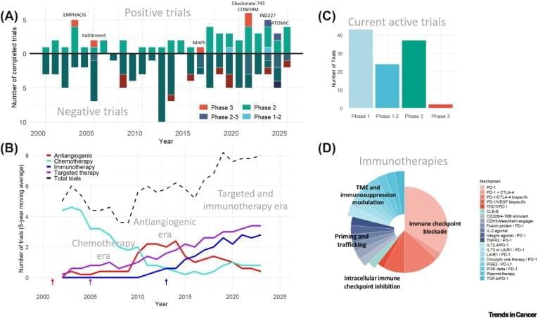
Mesothelioma is a rare cancer that has seen few incremental improvements in survival over the past two decades. However, a significantly improved understanding of the underlying biology has led to new therapeutic advances with the potential to improve clinical outcomes. In this review, we take a snapshot of the current systemic therapy research landscape, with our goal to forecast the trajectory of drug development for mesothelioma over the next half-decade. In our current census, we identify 106 active trials including systemic therapies: 20 (19%) are molecularly targeted, 26 (25%) include immunomodulation, and 12 (11%) combining immunotherapy with antiangiogenic therapies. Collectively, the landscape of therapeutic innovation for mesothelioma is expanding, bringing hope that improvements in life expectancy may follow.