Kilili, H., Padilla-Morales, B., Castillo-Morales, A. et al. Sci Rep 15, 15,087 (2025). https://doi.org/10.1038/s41598-025-98786-3
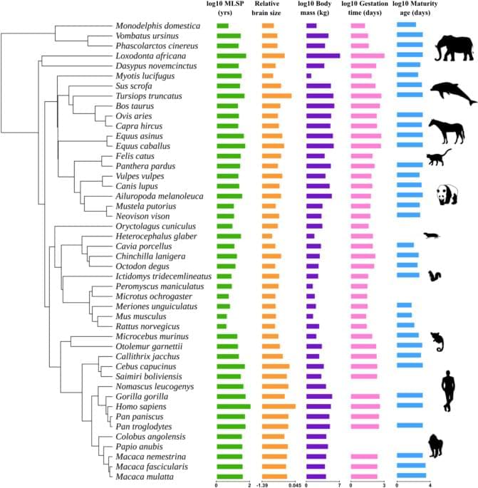
Kilili, H., Padilla-Morales, B., Castillo-Morales, A. et al. Sci Rep 15, 15,087 (2025). https://doi.org/10.1038/s41598-025-98786-3

The field of nanotechnology is still in its nascent stages, but recent innovations are increasingly making this science fiction world of tiny robots into a reality. New breakthrough research from a team at Caltech has demonstrated the ability of a robot made of a single strand of DNA to explore a molecular surface, pick up targeted molecules, and move them to another designated location.
“Just like electromechanical robots are sent off to faraway places, like Mars, we would like to send molecular robots to minuscule places where humans can’t go, such as the bloodstream,” says Lulu Qian, co-author on the paper. “Our goal was to design and build a molecular robot that could perform a sophisticated nanomechanical task: cargo sorting.”
Previous work by a variety of researchers has successfully demonstrated the creation of such DNA robots, but this is the first time they have been shown to pick up and transport specific molecules.

In the absence of air, microorganisms produce hydrogen using an enzyme called [FeFe]-hydrogenase, one of the most efficient hydrogen-producing biocatalysts known and a promising tool for green hydrogen energy. However, these enzymes are rapidly destroyed when exposed to air, which has so far limited their industrial use.
Now, joint efforts led by scientists from the Photobiotechnology group and the Center for Theoretical Chemistry at Ruhr University Bochum, Germany, have isolated a new type of oxygen-stable [FeFe]-hydrogenase and revealed its “tricks” for this oxygen-stability.
The results are published in the Journal of the American Chemical Society.

NASA’s James Webb Space Telescope (JWST) utilizes mid-infrared spectroscopy to precisely analyze molecular components such as water vapor and sulfur dioxide in exoplanet atmospheres. The key to this analysis, where each molecule exhibits a unique spectral “fingerprint,” lies in highly sensitive photodetector technology capable of measuring extremely weak light intensities.
Recently, KAIST researchers have developed an innovative photodetector capable of detecting a broad range of mid-infrared spectra, garnering significant attention. A research team led by Professor SangHyeon Kim from the School of Electrical Engineering has developed a mid-infrared photodetector that operates stably at room temperature, marking a major turning point for the commercialization of ultra-compact optical sensors.
The work is published in the journal Light: Science & Applications.

X-ray imaging is indispensable in medical diagnostics and material characterization. To generate an image, a detector converts X-rays that pass through the object into electrical signals. Higher detector sensitivity enables lower radiation doses, which is particularly important in medical applications.
Currently used X-ray detectors consist of inorganic compounds of elements with medium to high atomic numbers. In recent years, inorganic perovskite compounds have also been tested as X-ray detectors with very good results.
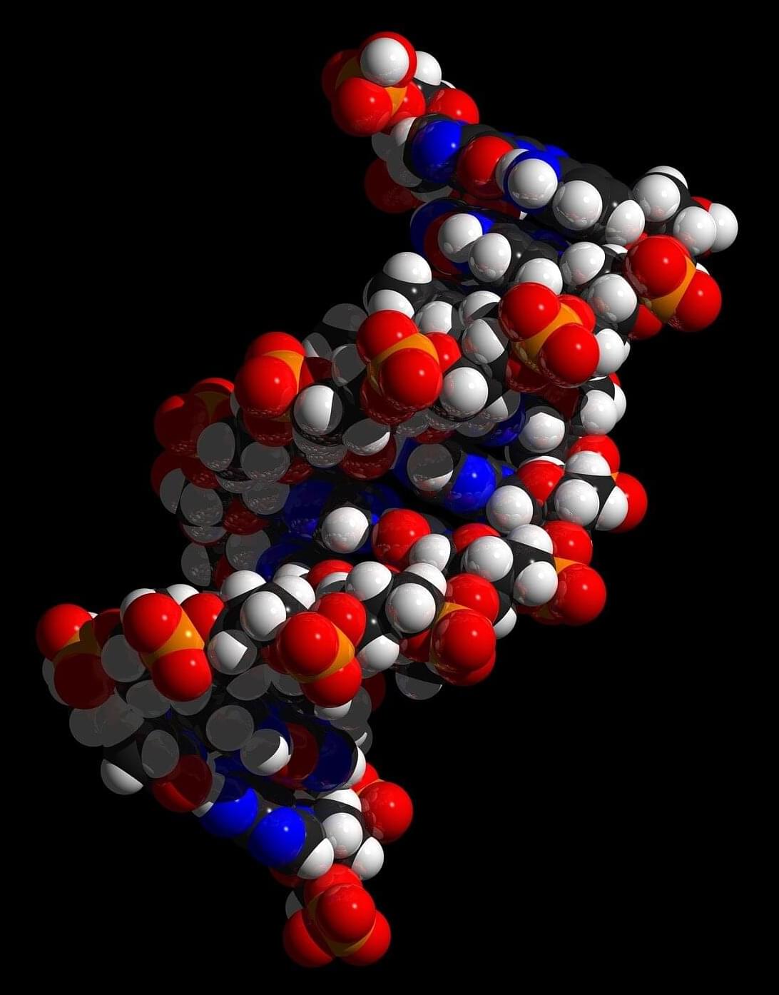
Most biochemistry labs that study DNA isolate it within a water-based solution that allows scientists to manipulate DNA without interacting with other molecules. They also tend to use heat to separate strands, heating the DNA to more than 150°F, a temperature a cell would never naturally reach. By contrast, in a living cell DNA lives in a very crowded environment, and special proteins attach to DNA to mechanically unwind the double helix and then pry it apart.
“The interior of the cell is super crowded with molecules, and most biochemistry experiments are super uncrowded,” said Northwestern professor John Marko. “You can think of extra molecules as billiard balls. They’re pounding against the DNA double helix and keeping it from opening.”

What if the secret to longevity wasn’t in the mind or the gut — but in the heart?
Speaking at the inaugural New York Times Well Festival on Wednesday, psychiatrist and researcher Dr. Robert Waldinger announced he and his team were “shocked” by “the biggest predictor of who was going to live long and stay healthy.”
Waldinger, the director of the Harvard Study of Adult Development — the longest-running scientific study of adult life — revealed the predictor was “how connected you were to other people and particularly the warmth of your connection to other people.”
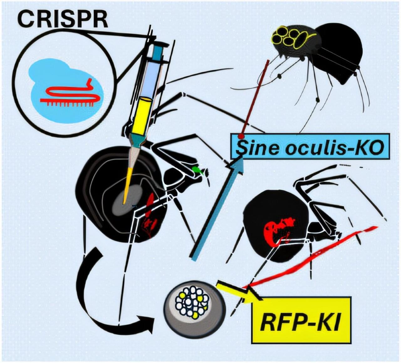
The University of Bayreuth’s Biomaterials research group has, for the first time, successfully applied the CRISPR-Cas9 gene-editing tool to spiders. Following the genetic modification, the spiders produced red fluorescent silk.
The findings of the study have been published in the journal Angewandte Chemie.
Spider silk is one of the most fascinating fibers in the field of materials science. In particular, its dragline thread is extremely tear-resistant, while also being elastic, lightweight and biodegradable. If scientists succeed in influencing spider silk production in vivo—in a living animal—and thereby gain insights into the structure of the dragline thread, it could pave the way for the development of new silk functionalities for a wide range of applications.

An annual blood test could prevent around half of cancer cases from reaching an advanced stage, new research suggests.
Scientists are currently trying to determine the effectiveness of simple blood tests in detecting cancer before symptoms appear, and whether such early detection improves survival rates.
The NHS is currently trialling such tests, including the Galleri test and miONCO-Dx test, with experts predicting a nationwide rollout of a universal cancer screening program within the next decade.