Researchers have identified a biomarker in spinal fluid that can detect Parkinson’s disease in its early stages with over 90% accuracy.

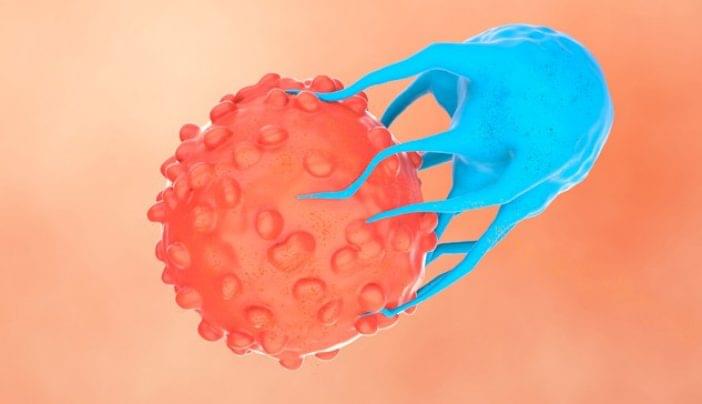
For decades, researchers have been exploring ways to harness the power of the immune system to treat cancer. One breakthrough is cell therapy, often called ‘living drugs.’ This is a form of immunotherapy that uses immune cells from a patient or a healthy donor. With advanced engineering techniques, scientists enhance these cells to recognize better and attack cancer.
“During the late 1980s and 1990s, cancer researchers started exploring ways to advance immunotherapy by transferring immune cells into a patient to attack cancer cells,” says stem cell transplant and cellular therapy specialist Hind Rafei, M.D. “They recognized that immune cells found inside tumors could help destroy cancer cells, leading to the development of one of the earliest forms of cell therapy — tumor-infiltrating lymphocytes (TILs).”
Cell therapy is a form of immunotherapy that uses immune cells from a patient or a healthy donor to treat cancer. Learn about the types of cell therapy from stem cell transplant and cellular therapy specialist Hind Rafei, M.D.

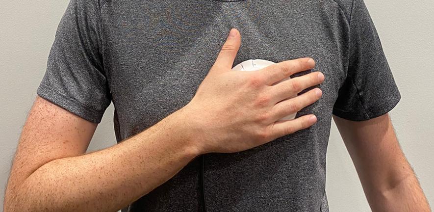
Researchers have developed a handheld device that could potentially replace stethoscopes as a tool for detecting certain types of heart disease.
The researchers, from the University of Cambridge, developed a device that makes it easy for people with or without medical training to record heart sounds accurately. Unlike a stethoscope, the device works well even if it’s not placed precisely on the chest: its larger, flexible sensing area helps capture clearer heart sounds than traditional stethoscopes.
The device can also be used over clothing, making it more comfortable for patients – especially women – during routine check-ups or community heart health screening programmes.
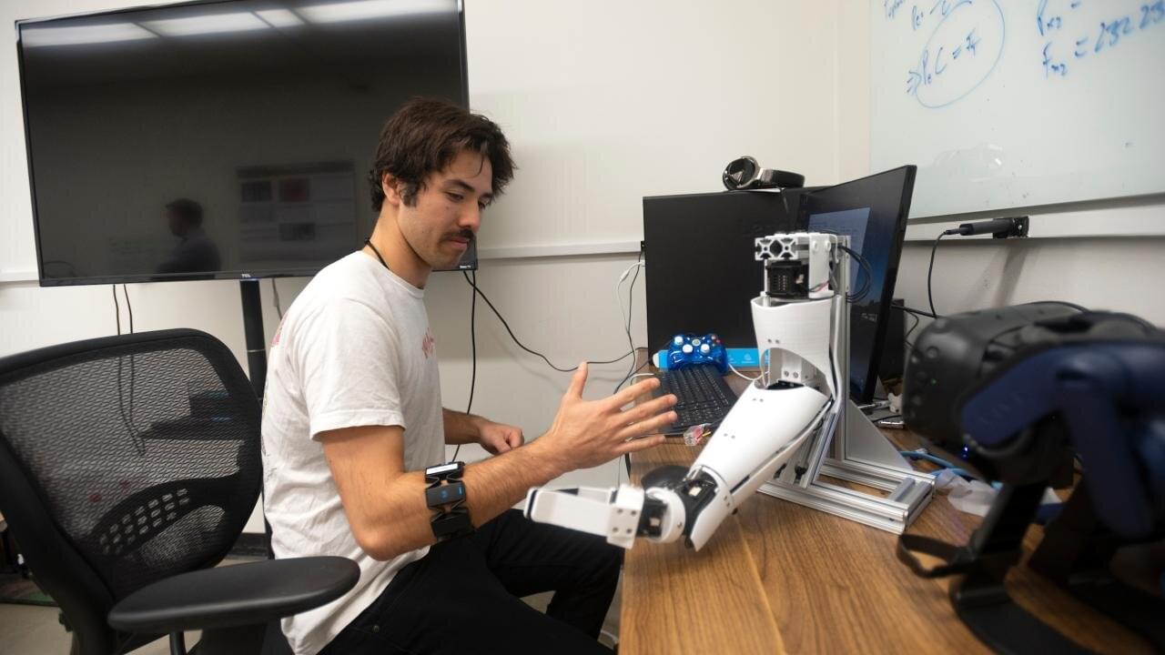
Combining two different kinds of signals could help engineers build prosthetic limbs that better reproduce natural movements, according to a new study from the University of California, Davis. The work, published April 10 in PLOS One, shows that a combination of electromyography and force myography is more accurate at predicting hand movements than either method by itself.
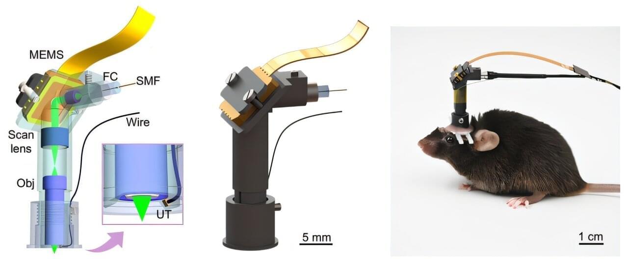
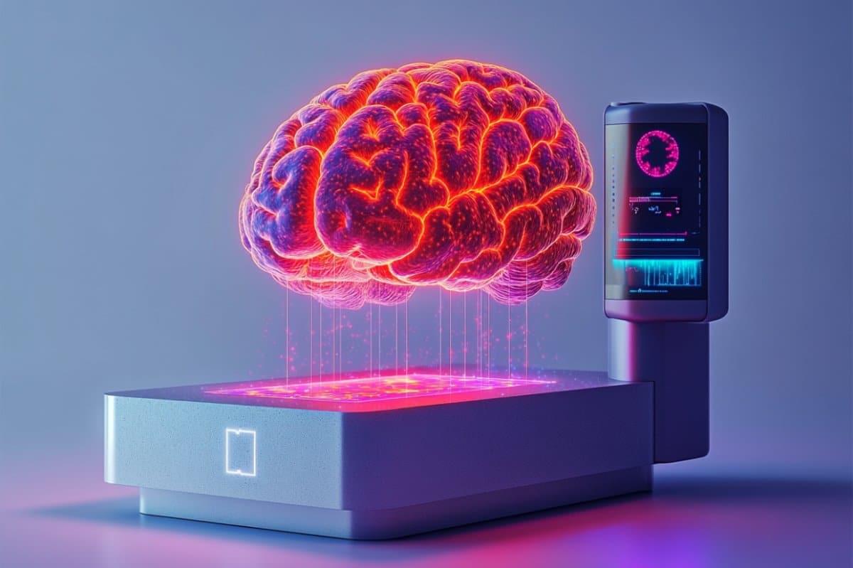
In a study published in Science Advances, a research team led by Prof. Liu Chengbo from the Shenzhen Institutes of Advanced Technology (SIAT) of the Chinese Academy of Sciences developed a 1.7-gram head-mounted microscope. This innovative device can simultaneously capture neural activity and cerebral hemodynamics in freely moving mice, providing a novel tool for exploring neurovascular coupling (NVC) in the brain and studying neurological disorders.
NVC represents the close temporal and regional relationship between neural activity and cerebral blood flow and oxygenation. When neural activity is initiated in a specific brain region, the metabolic demand within that region increases, leading to enhanced blood flow to supply greater amounts of oxygen and glucose, thus meeting the elevated metabolic needs of neurons. Conversely, if pathological changes hinder cerebral vessels’ capacity to provide sufficient oxygen and energy, the functional activity of the corresponding brain region will be significantly affected.
Conventional NVC imaging techniques face challenges in achieving simultaneous in vivo high spatiotemporal resolution of neuronal activity and cerebral blood flow. This makes it difficult to accurately capture the dynamic relationship between neural activity and local hemodynamic changes. In addition, most existing studies use head-fixed setups which do not reflect true neurovascular coupling during natural animal behaviors.
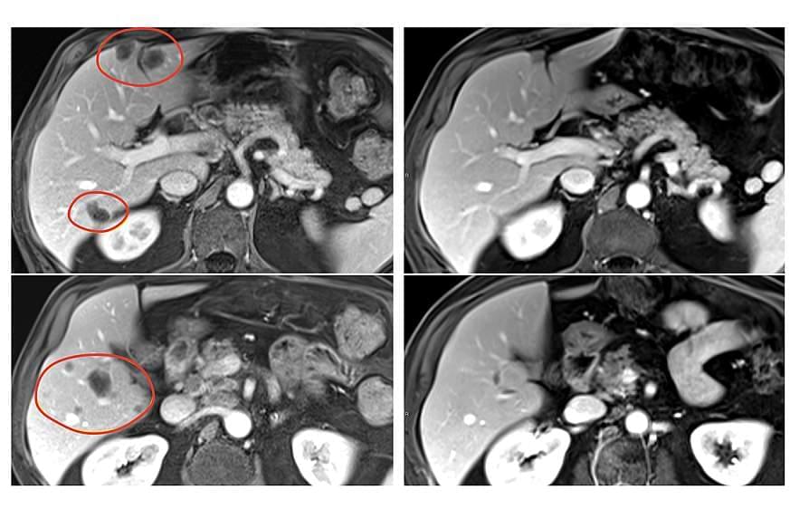
A research team has developed an innovative biomimetic dual-mode magnetic resonance imaging (MRI) nanoprobe for detecting early-stage liver fibrosis in non-alcoholic fatty liver disease (NAFLD).
The work, using the Steady High Magnetic Field Facility (SHMFF), was recently published in Advanced Science. The team was led by Prof. Wang Junfeng at the High Magnetic Field Laboratory, the Hefei Institutes of Physical Science of the Chinese Academy of Sciences.
NAFLD is a growing global health concern with even higher rates among individuals with obesity or type 2 diabetes. Detecting liver fibrosis early, before it becomes irreversible, is crucial for timely intervention and treatment. While MRI is a promising non-invasive tool for identifying liver fibrosis, traditional imaging techniques often lack the sensitivity to catch early-stage changes. Conventional contrast agents either face safety concerns or fail to target fibrotic tissues specifically.