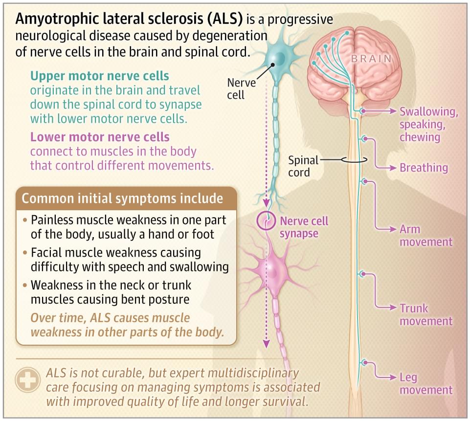
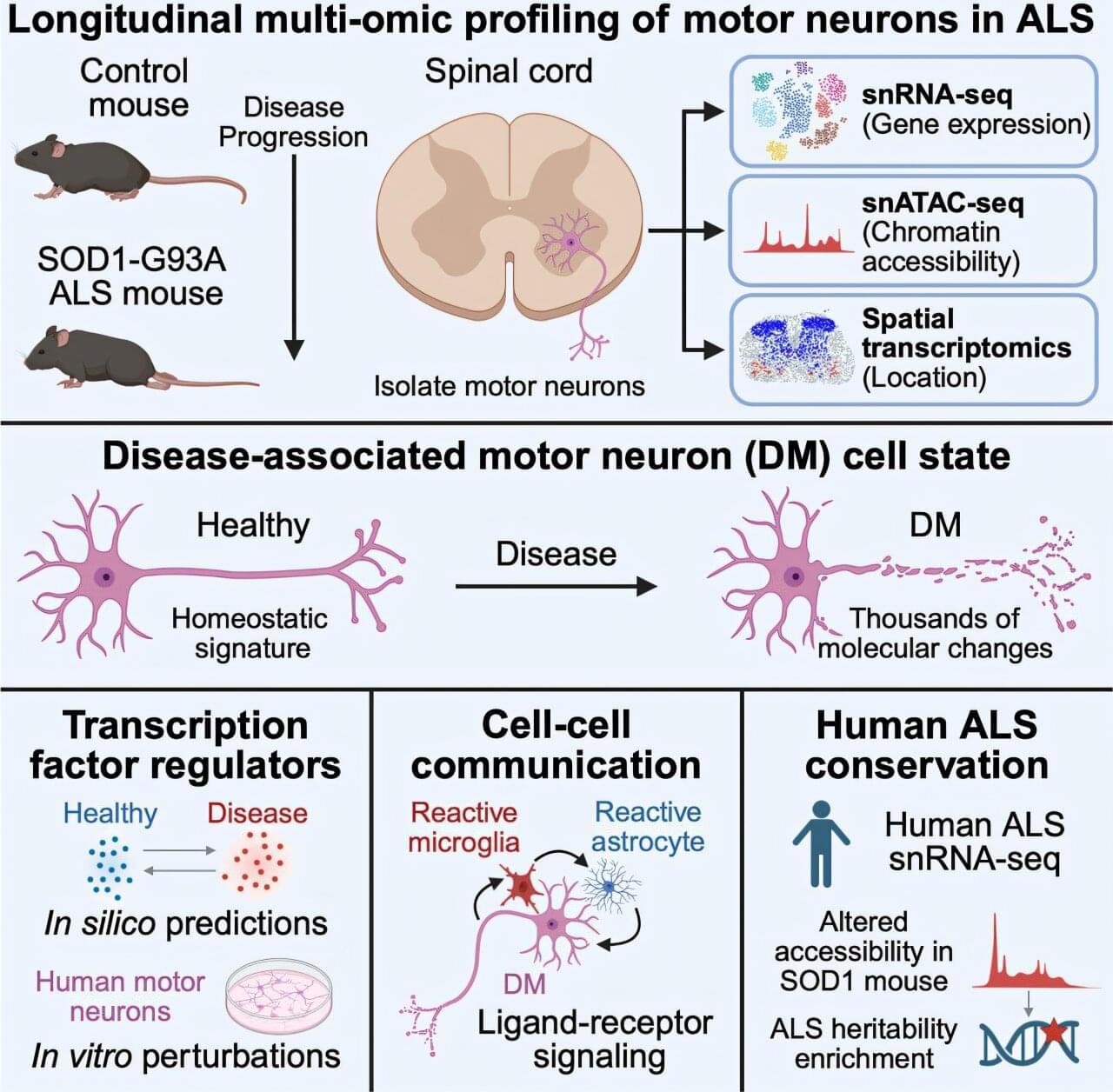
Toggle light / dark theme

What if a simple vial of synthetic blood could save millions of lives? From cutting-edge laboratories to the frontlines of disaster zones, scientists are revolutionizing medicine with the development of artificial blood. In this episode of Beyond the Veil, we take you on a journey into the world of groundbreaking innovations like ErythroMer, a shelf-stable synthetic red blood cell, and lab-grown blood cells that could transform healthcare as we know it.

Join us as we explore:
🔬 The intricate science behind replicating blood\’s vital functions.
💡 How Dr. Allan Doctor and his team are making synthetic red blood cells a reality with ErythroMer.
đŸ©ž The NHS’s revolutionary trial of lab-grown red blood cells.
🌍 The potential to save lives in remote areas, on battlefields, and even during space exploration.
🚀 The hurdles, breakthroughs, and future implications of artificial blood research.

💉 This isn\’t just a story about science; it\’s a tale of perseverance, hope, and the determination to rewrite the rules of medicine.

Sources and Research Cited in this Episode:

Identifying and delineating cell structures in microscopy images is crucial for understanding the complex processes of life. This task is called “segmentation” and it enables a range of applications, such as analyzing the reaction of cells to drug treatments, or comparing cell structures in different genotypes.

It was already possible to carry out automatic segmentation of those biological structures, but the dedicated methods only worked in specific conditions and adapting them to new conditions was costly. An international research team led by Göttingen University has now developed a method for retraining the existing AI-based software Segment Anything on over 17,000 with over 2 million structures annotated by hand.

The new model is called Segment Anything for Microscopy and it can precisely segment images of tissues, cells and similar structures in a wide range of settings. To make it available to researchers and medical doctors, they have also created ÎŒSAM, user-friendly software to “segment anything” in microscopy images. The work is published in Nature Methods.

Super cool paper where Jeppesen et al. discover and characterize a new type of large extracellular vesicle (EV) that they call blebbisomes! These blebbisomes have active mitochondria as well as other organelles (except nucleus), secrete and take up smaller EVs, and can reach sizes of up to 20 micrometers! #cellbiology #molecularbiology #biochemistry


Cells release a variety of 30-to 10,000-nm lipid-bilayer-enclosed extracellular vesicles (EVs) to facilitate cell-to-cell and cell-to-environment communication by packaging signalling molecules to avoid degradation1,2,3,4,5 and escape immune surveillance6,7,8,9. EVs may interact with target cells through contact between molecules on the EV surface with receptors on the cell surface to relay signals. In addition, modulation of recipient cell behavior may follow uptake of EVs cargo, including bioactive proteins, lipids and nucleic acids. EVs have emerged as important actors and agents of intercellular communication in normal cell biology and pathological conditions2,4,6.

Here, we identify blebbisomes, an exceptionally large functional EVs, that are actively released by human and mouse cells, remain motile independently of cells and have the capacity to both take up EVs and secrete exosomes and microvesicles. Blebbisomes are the largest type of EV described so far with an average diameter of 10 ”m but can be as large as 20 ”m, with an area commonly larger than 50 ”m2. After being released from motile cells, blebbisomes display marked contractility-dependent ‘blebbing’ behaviour. Both normal and cancer cells release blebbisomes that contain active, healthy, mitochondria further distinguishing them from other large EVs (lEVs) such as exophers10,11 and migrasomes12 that function in the removal of damaged mitochondria from cells under stress conditions. In addition, blebbisomes contain many other cellular organelles including endoplasmic reticulum (ER), Golgi apparatus, ribosomes, lysosomes, endosomes, multivesicular endosomes (MVEs) and autophagosomes/amphisomes, as well as cytoskeletal elements; however, they lack a definable nucleus.

Researchers from Cleveland Clinic’s Genome Center have outlined the pathway human herpes simplex virus-1 (HSV1) can use to contribute to Alzheimer’s disease in aging brains. In a report published in Alzheimer’s & Dementia, investigators also share two FDA-approved, commercially available drugs that reverse this pathway in a laboratory setting.

The findings are the first concrete evidence to support the previously controversial link between human herpesviruses (HHVs) and Alzheimer’s disease. Illustrating the potential for herpes to trigger dementia aids continued efforts to prevent and cure neurodegenerative disease, says senior author and Genome Center director Feixiong Cheng, Ph.D


For most people, contracting a is just an inconvenient or harmless fact of life. Many herpesviruses are individually present in a large percentage of people worldwide, meaning virtually every human being on earth is expected to contract at least three types of herpesviruses by adulthood. Some of these viruses don’t cause symptoms, while others only cause minor illnesses like mono or chickenpox. However, even after these illnesses subside, an infected individual still carries herpesviruses for the rest of their life, with only minor symptoms like occasional cold sores.

New findings cast light on the role of understudied DN3 memory B cells in cancer, and shows these cells correlate with progression and poor clinical outcomes in head and neck squamous cell carcinoma and melanoma.

📄


Tumor-associated extrafollicular B cells are phenotypically, locationally, and functionally distinct from germinal center–dependent memory B cells.

An AI-powered tool called MELD Graph is revolutionizing epilepsy care by detecting subtle brain abnormalities that radiologists often miss.

By analyzing global MRI data, the tool improves diagnosis speed, increases access to surgical treatment, and cuts healthcare costs. Though not yet in clinical use, it is already helping doctors identify operable lesions, offering hope to epilepsy patients worldwide.

AI Breakthrough in Epilepsy Detection.

There’s an arms race in medicine—scientists design drugs to treat lethal bacterial infections, but bacteria can evolve defenses to those drugs, sending the researchers back to square one. In an article published in the Journal of the American Chemical Society, a University of California, Irvine-led team describes the development of a drug candidate that can stop bacteria before they have a chance to cause harm.

“The issue with antibiotics is this crisis of antibiotic resistance,” said Sophia Padilla, a Ph.D. candidate in chemistry and lead author of the new study. “When it comes to antibiotics, can evolve defenses against them—they’re becoming stronger and always getting better at protecting themselves.”

About 35,000 people in the U.S. die each year from from pathogens like Staphylococcus, while about 2.8 million people suffer from bacteria-related illnesses.

Researchers at the Technical University of Munich (TUM) have invented an entirely new field of microscopy called nuclear spin microscopy. The team can visualize magnetic signals of nuclear magnetic resonance with a microscope. Quantum sensors convert the signals into light, enabling extremely high-resolution optical imaging.

Magnetic resonance imaging (MRI) scanners are known for their ability to look deep into the human body and create images of organs and tissues. The new method, published in the journal Nature Communications, extends this technique to the realm of microscopic detail.

“The used make it possible to convert signals into optical signals. These signals are captured by a camera and displayed as images,” explains Dominik Bucher, Professor of Quantum Sensing and researcher at the Cluster of Excellence Munich Center for Quantum Science and Technology (MCQST).

Porcine reproductive and respiratory syndrome virus (PRRSV) infection in pigs is economically devastating for the global swine industry. The viral infection leads to reproductive disorder in sows and respiratory problems in infected newborn and growing pigs.

Unfortunately, high genetic variability of the virus and differing disease-causing strength or virulence hinders vaccine development and complicates disease management. Not much is known about the factors contributing to viral disease severity or the anti-viral immune responses.

Dr. Jun-Mo Kim, Associate Professor at the Department of Animal Science and Technology, Chung-Ang University, Korea, has focused his research efforts on filling this gap in understanding.