Solriamfetol, a new treatment for ADHD, showed a 45% reduction in symptoms in a Phase 3 trial. This treatment targets dopamine and norepinephrine reuptake and offers a safer alternative for those who don’t respond well to traditional stimulants.

Parkinson’s doesn’t just affect movement and the brain—it may also impact the heart, according to new research from the University of Surrey. Scientists from Surrey’s School of Veterinary Medicine suggest that targeting a key protein outside of the brain could help manage Parkinson’s-related heart issues.
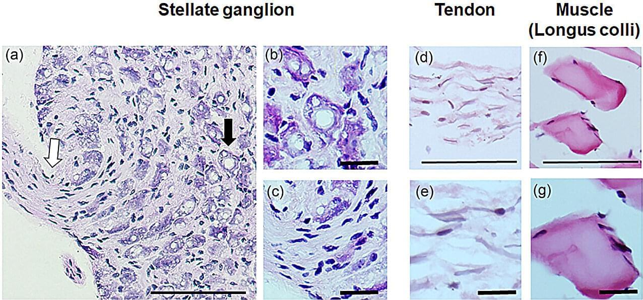
In a study published in Experimental Physiology, Surrey researchers studied mouse models and found a harmful buildup of the alpha-synuclein protein, which is associated with Parkinson’s disease, in a nerve cluster near the heart (the stellate ganglia). These nerves are part of the autonomic nervous system, which controls heart rate and rhythm.
Researchers found that 27% of neurons in the nerve cluster contained aggregated alpha-synuclein, forming similar toxic clumps seen in the brains of Parkinson’s patients. This finding suggests that Parkinson’s could disrupt heart function, not just movement and brain activity.

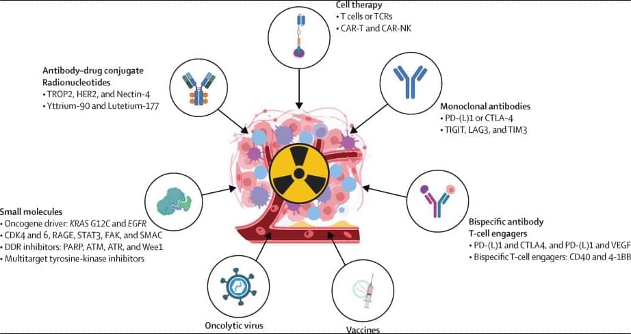
Over the past few decades, breakthroughs in cancer biology at the molecular level have revolutionised cancer treatment. Enhanced precision in radiotherapy has not only reduced patient side-effects, but also enabled the delivery of high-dose stereotactic extracranial irradiation with unprecedented accuracy. Simultaneously, the number of medical therapies available for clinical care continues to grow. Despite the progress made with combined chemoradiotherapy, only a few drug–radiotherapy combinations have received clinical approval, leaving a vast landscape of untapped opportunities for basic, translational, and clinical research, particularly in early-phase drug–radiotherapy trials.
Join us on Patreon! https://www.patreon.com/MichaelLustgartenPhD
Discount Links/Affiliates:
Blood testing (where I get the majority of my labs): https://www.ultalabtests.com/partners/michaellustgarten.
At-Home Metabolomics: https://www.iollo.com?ref=michael-lustgarten.
Use Code: CONQUERAGING At Checkout.
Clearly Filtered Water Filter: https://get.aspr.app/SHoPY
Epigenetic, Telomere Testing: https://trudiagnostic.com/?irclickid=U-s3Ii2r7xyIU-LSYLyQdQ6…M0&irgwc=1
Use Code: CONQUERAGING
NAD+ Quantification: https://www.jinfiniti.com/intracellular-nad-test/
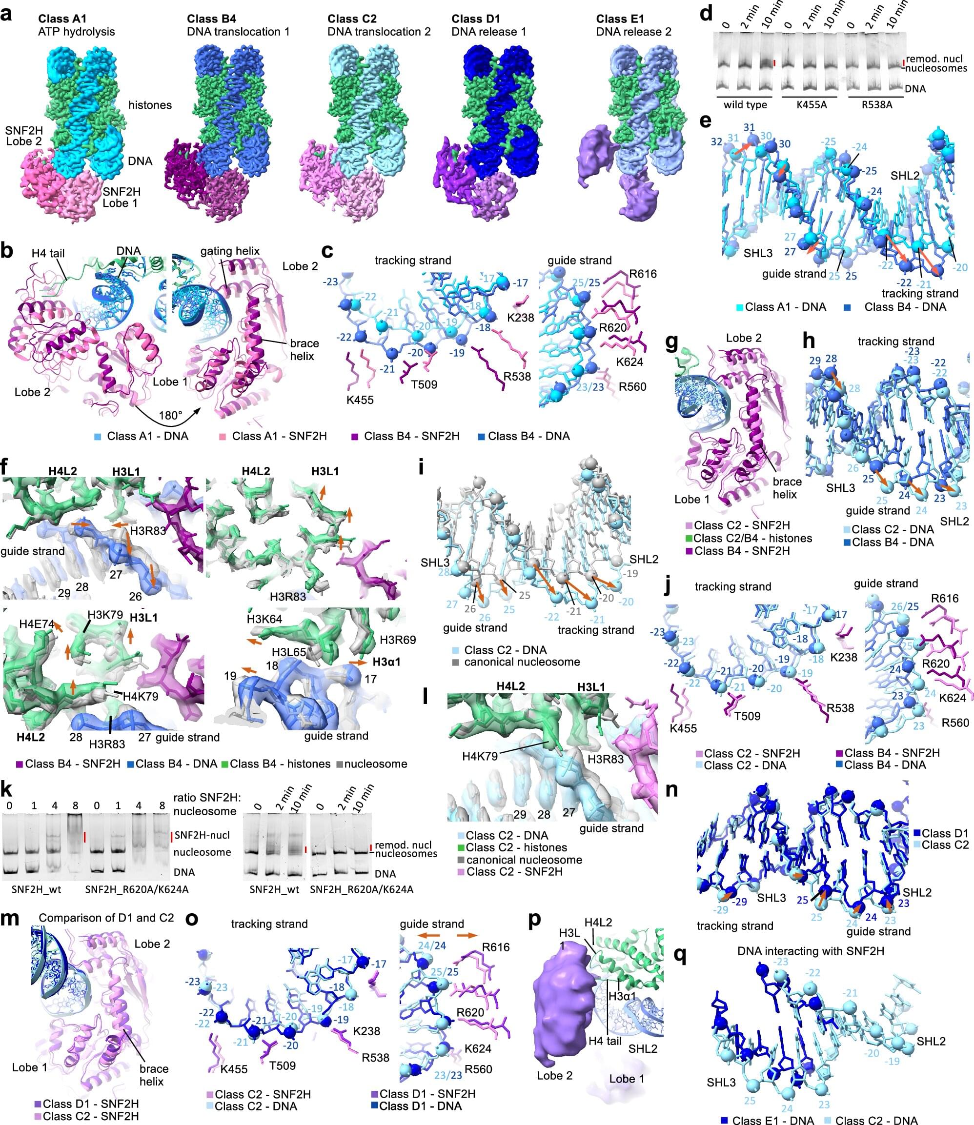
Chromatin remodeling plays a vital role in gene regulation, affecting how DNA is accessed. Disruptions in this process can also lead to cancer and other diseases.
To better understand how chromatin remodeling works, scientists at St. Jude Children’s Research Hospital used cryo–electron microscopy (cryo-EM) to obtain fine structural details of a human chromatin remodeler in action.
The researchers captured 13 structures that together offer a comprehensive view of how the remodeling enzyme SNF2H works, offering insights that are likely shared across other such enzymes. The work was published today in Cell Research.
Polymer-coated nanoparticles loaded with therapeutic drugs show significant promise for cancer treatment, including ovarian cancer. These particles can be targeted directly to tumors, where they release their payload while avoiding many of the side effects of traditional chemotherapy.
Over the past decade, MIT Institute Professor Paula Hammond and her students have created a variety of these particles using a technique known as layer-by-layer assembly. They’ve shown that the particles can effectively combat cancer in mouse studies.
To help move these nanoparticles closer to human use, the researchers have now come up with a manufacturing technique that allows them to generate larger quantities of the particles, in a fraction of the time.

Differences in the distribution of certain proteins and markers in the brain may explain why some people first experience vision changes instead of memory loss in Alzheimer’s disease, finds a new study by UCL researchers.
Posterior cortical atrophy (PCA) is a rare form of Alzheimer’s disease that, rather than causing problems with memory, leads to difficulties with reading, navigating, and recognizing objects. Studies suggest that one in 10 patients with Alzheimer’s disease has a form which is visual, rather than memory-led.
As well as presenting with unusual symptoms, individuals with PCA typically develop symptoms younger than most people with Alzheimer’s disease, with onset usually in their 50s and 60s.
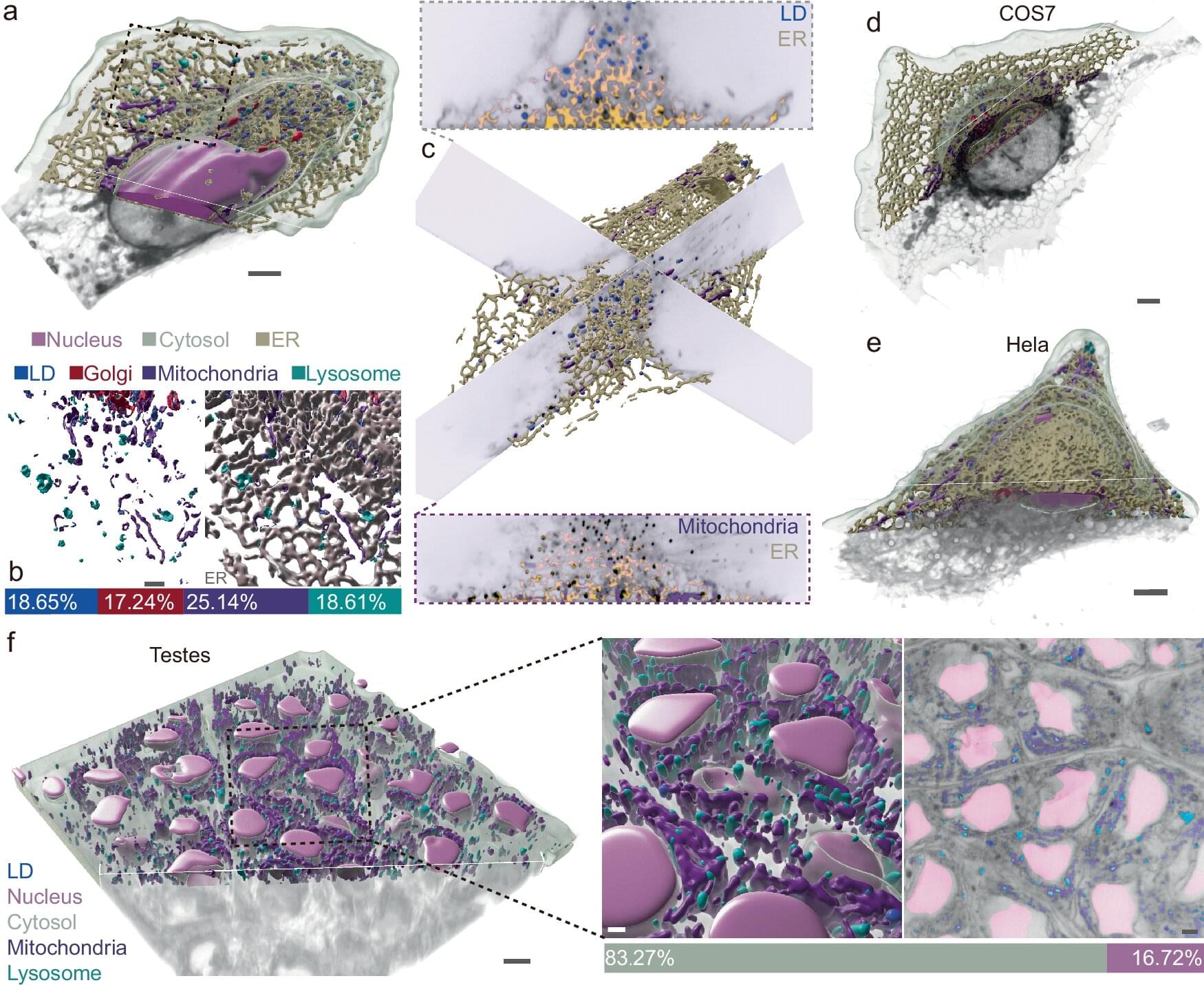
A breakthrough in imaging technology promises to transform our understanding of the inner workings of living cells, and provide insights into a wide range of diseases.
The study, recently published in the journal Nature Communications, unveils an innovative approach that combines super-resolution imaging with artificial intelligence and deep learning to reveal subcellular structures and dynamics. It was led by researchers from Peking University, Ningbo Eastern Institute of Technology and the University of Technology Sydney.
“It’s like taking an airplane over a city at night and watching all the live interactions,” said UTS Distinguished Professor Dayong Jin. “This cutting-edge technology will open new doors in the quest to understand the intricate world within our cells.”