W/ Dr. Andy Norman of Carnegie Mellon University. The prospects of cognitive immunology which posits mental immune systems & proceeds to investigate their functioning.


Alzheimer’s disease (AD), the most prevalent neurodegenerative disorder, continues to pose significant challenges despite advances in anti-amyloid therapies. New research from Harvard Medical School and Washington University School of Medicine, published in Science Translational Medicine, has unveiled a novel therapeutic approach: the use of inhaled xenon gas to modulate microglia and ameliorate disease progression in mouse models of AD.
How Xenon Targets Microglia
Microglia, the brain’s resident immune cells, play a dual role in neurodegeneration. While they can clear amyloid-beta (Aβ) plaques and damaged neurons, chronic activation leads to neuroinflammation, contributing to disease progression. Xenon gas, an inert anaesthetic, penetrates the blood-brain barrier and appears to modulate microglia to adopt a “pre-neurodegenerative microglia” (pre-MGnD) state.
Researchers have identified a key pathway that links how neurons send signals to each other, or synaptic activity, to the expression of genes necessary for long-term changes in the brain, providing crucial insights into the molecular processes underlying memory formation.
“These findings illuminate a critical mechanism that connects local synaptic activity to the broader gene expression changes necessary for learning and memory,” said Mark Dell’Acqua, professor of pharmacology at the University of Colorado Anschutz Medical Campus and senior author of the study. “This paper is mainly a basic science finding of a fundamental process of what nerve cells do. Understanding this relay system not only enhances our knowledge of brain function but could also better inform therapeutic treatments for cognitive disorders.”
The nucleus where the genes that modify neuron function are controlled is a long distance away from where neurons receive input from their synapses, which are located in distant dendrites that extend like branches from the trunk of a tree. This research focuses on the cAMP-response element binding protein (CREB), a transcription factor known to regulate genes vital for dynamic changes at synapses which is essential for neuronal communication. Despite CREB’s well-documented role in supporting learning and memory, the exact mechanisms leading to CREB activation during neuronal activity remain unclear.
Using advanced microscopy techniques, graduate student Katlin Zent in Dr. Dell’Acqua’s research group revealed a crucial relay mechanism involving the activation of receptors and ion channels generating calcium signals that rapidly communicates from synapses in remote dendrite branches to the nucleus in the neuron cell body.
“Going forward, this research will enable us to better examine the way these pathways are utilized in different disease states,” said Dell’Acqua. “We could see exactly what parts of this new mechanism are interfered with and where, giving us a better idea of how this pathway affecting learning and memory is impacted. This research highlights potential targets for interventions aimed at conditions like Alzheimer’s disease and other memory-related disorders.”
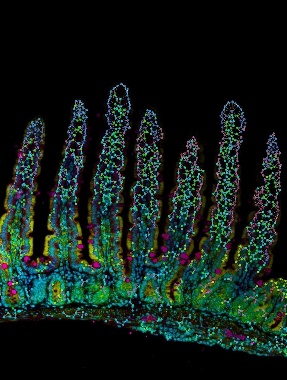
The human immune system is like an army of specialized soldiers (immune cells) each with a unique role to play in fighting disease. In a new study published in Nature, led by scientists at the Allen Institute, La Jolla Institute for Immunology, and UC San Diego, researchers reveal how cells known as tissue-resident memory CD8 T cells, play unique and specialized roles based on where they are located within the small intestine.
Tissue-resident memory cells provide a local first line of defense against re-infection and call for “backup” from other immune cells and are also critical for maintaining peace in a tissue exposed to many outside pathogens.
This discovery sheds light on how tissue-resident memory CD8 T cells adapt to their location in the body, ensuring a coordinated and effective immune response and how microenvironments and cellular interactions shape this location-specific adaptation. Ultimately, location matters, and this understanding could also lead to improved immunotherapy and vaccines.
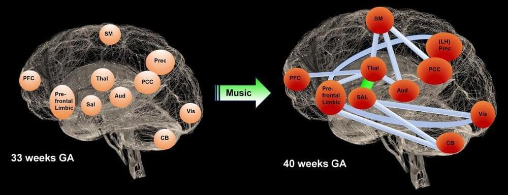
Certain melodies promote brain development in premature infants. For several years, a team of scientists have observed this phenomenon. They now know more precisely which areas of the brain react over time.
Premature infants are more likely to suffer from attention and emotional regulation disorders. For more than a decade, a team has been investigating an unexpected solution to prevent these problems: music. Scientists at the Geneva University Hospitals (HUG) exposed several cohorts of infants born at an average of 29 weeks to music.
Several of their publications, which have been widely covered in the media, underline the potential of this approach. The team’s latest study demonstrates that music boosts cerebral connectivity in the areas of the brain usually affected in preterm infants.
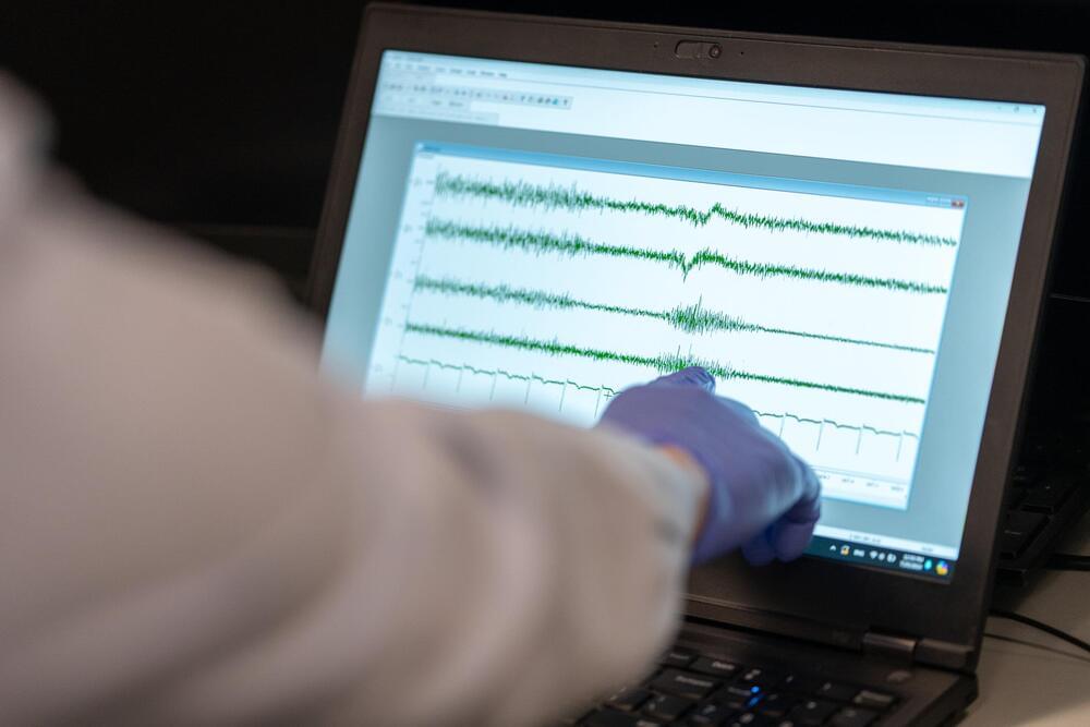
From the ancient Egyptians’ use of electric fish to treat headaches to the invention of pacemakers to regulate heart rhythms in the 1950s, the field of bioelectronic medicine—which makes use of electrical signals instead of drugs to diagnose and treat disease—has advanced and has started to come into its own. Where is the field now? And what are the most promising opportunities for life-changing new therapies and diagnostics?
New research led by Imanuel Lerman, head of the Lerman Lab of the UC San Diego Qualcomm Institute and UC San Diego School of Medicine Department of Anesthesiology, as well as the VA Center of Excellence for Stress and Mental Health, provides some answers.
“This paper is intended to be a roadmap to the future of the biomedicine field,” Lerman said. “We’re putting a flagpole in the ground and saying, ‘This is what we’re planning to do, and this is the story behind it.’ That’s why there are 180 references. We want to make sure that everybody has the resources they may need to be able to understand and read deeper if they want to.”
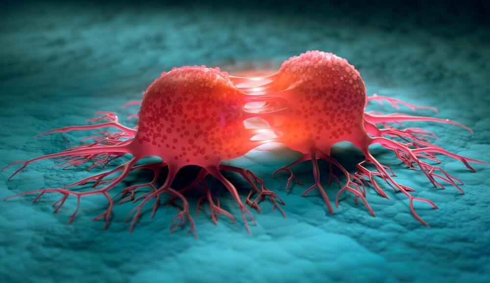
Researchers have found that kinesin family member 9 (KIF9), a protein that diminishes with aging, is instrumental in allowing cells to consume harmful proteins and fights Alzheimer’s in a mouse model.
Consuming amyloids before they become a problem.

The ability to regulate one’s own food intake is essential to the survival of both humans and other animals. This innate ability ensures that the body receives the nutrients it needs to perform daily activities, without significantly exceeding calorie intake, which could lead to health problems and metabolic disorders.
Past neuroscience studies suggest that the regulation of food intake is supported by specific regions in the brain, including the hypothalamus and caudal nucleus of the solitary tract (cNTS), which is part of the brainstem. This key region in the brainstem is known to integrate sensory signals originating from the gut and then transform them into adaptive feeding behaviors.
While previous research has highlighted the key role of the cNTS in food intake regulation, the unique contribution of the different neuron subtypes within this brainstem region and the mechanisms by which they regulate feeding remain poorly understood. Better understanding these neuron-specific mechanisms could help to devise more effective therapeutic interventions for obesity and eating disorders.