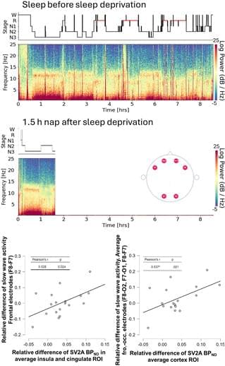
Toggle light / dark theme

Key alterations discovered in the cerebral cortex of people with psychosis

Researchers at the University of Seville have analyzed alterations in the cerebral cortex in people suffering from psychosis. Their findings show that psychosis does not follow a single trajectory, but rather its evolution depends on a complex interaction between brain development, symptoms, cognition and treatment. The authors therefore emphasize the need to adopt more personalized approaches that take individual differences into account in order to better understand the disease and optimize long-term therapeutic strategies.

Psychosis is a set of symptoms—such as hallucinations and delusions—that are common in schizophrenia and involve a loss of contact with reality. From their first manifestation, known as the first psychotic episode, these symptoms can appear and evolve in very different ways between individuals, thus making schizophrenia a particularly complex disorder.

The results of the study show that, at the time of the first episode, people with psychosis present a reduction in cortical volume, which is particularly marked in regions with a high density of serotonin and dopamine receptors, key neurotransmitters in both the pathophysiology of psychosis and the mechanism of action of antipsychotics. The data also suggest that both neurons and other brain cells involved in inflammatory and immunological processes may play an important role in the disease.

One stem cell generates 14 million tumor-killing NK cells in major cancer breakthrough

Scientists in China have unveiled a breakthrough way to mass-produce powerful cancer-fighting immune cells in the lab. By engineering early-stage stem cells from cord blood—rather than trying to modify mature natural killer (NK) cells—they created a streamlined process that generates enormous numbers of highly potent NK cells, including CAR-equipped versions designed to hunt specific cancers.

Symbiotic bacteria in planthoppers break record for smallest non-organelle genome ever found

Many insects rely on heritable bacterial endosymbionts for essential nutrients that they cannot get through their diet. A new study, published in Nature Communications, indicates that the genomes of these symbiotic bacteria often shrink over time. Some of these bacteria, which live inside certain insect cells, have lost so many genes that they have broken the record for the tiniest genome ever found—almost blurring the lines between organelle and bacteria.

Endosymbiotic relationships are common in many insects, and in sap-sucking insects, like planthoppers and cicadas, they are essential for the insect’s survival. Because the sap of plants does not typically contain certain amino acids or vitamins, the insect must get them another way. Over hundreds of millions of years, these insects have co-evolved with bacteria that provide these additional nutrients.

Sulcia and Vidania are two examples of bacterial endosymbionts, which have co-evolved with planthoppers for more than 260 million years. These bacteria live in specialized cells within the planthopper abdomen. The new study has found that, along with their hosts, these endosymbionts have evolved—or devolved—in some unexpected ways.

Scientists find hundreds of genes that behave like light switches

If cancer’s genetic off switch is found then even Covid 19s off switch could essentially be found aswell.


Gene expression, where cells use the genetic information encoded in DNA to produce proteins, has been thought of as a dimmer light.

How much a particular gene gets expressed continually rises and falls, depending on the needs of a cell at any given time. It’s like adjusting the lighting of a room until it’s just right for your mood.

But University at Buffalo researchers have shown that a considerable portion of a human’s roughly 20,000 genes express more like your standard light switch — fully on or fully off.

Anti-aging effect of Hedgehog signaling

Aging weakens the body’s ability to maintain balance and repair damage, increasing vulnerability to disease. This study reveals that the Hedgehog (Hh) signaling pathway plays a crucial role in preserving tissue integrity and regenerative capacity. Using animal models, researchers found that activating Hh signaling in multiple tissues such as the liver and brain enhances tissue repair and mitigates age-related functional decline. These findings suggest that targeting Hh signaling could be a promising strategy to promote healthy aging by enhancing regeneration and alleviating age-related dysfunction.

This summary was initially drafted using artificial intelligence, then revised and fact-checked by the author.

A PI3Kδ-Foxo1-FasL signaling amplification loop rewires CD4+ T cell signaling and differentiation

Dominic P. Golec, Pamela L. Schwartzberg and colleagues (National Institute of Allergy and Infectious Diseases (NIAID)) describe a PI3 Kinase-Foxo1-FasL signaling circuit that promotes amplified signaling and rewires transcriptional and epigenetic programs driving IFN-γ and altered T helper cell differentiation in CD4+ T cells from mice expressing an activating mutant of phosphoinositide 3-kinase delta.

Immunodeficiency LymphocyteBiology


While inputs regulating CD4+ T helper (Th) cell differentiation are well defined, the integration of downstream signaling with transcriptional and epigenetic programs that define Th lineage identity remains incompletely resolved. PI3K signaling is a critical regulator of T cell function; activating mutations affecting PI3Kδ result in an immunodeficiency with multiple T cell defects. Using mice expressing activated PI3Kδ, we found aberrant expression of proinflammatory Th1 signature genes under Th2-inducing conditions, both in vivo and in vitro. This dysregulation was driven by a PI3Kδ-IL-2-Foxo1 signaling amplification loop, fueling Foxo1 inactivation, loss of Th2 lineage restriction, and extensive epigenetic reprogramming. Surprisingly, ablation of Fasl, a Foxo1-repressed gene, normalized both Th2 differentiation and TCR signaling. BioID and imaging revealed Fas interactions with TCR signaling components, which were supported by Fas-mediated potentiation of TCR signaling that could occur in the absence of FADD. Our results highlight Fas-FasL signaling as a critical intermediate in phenotypes driven by activated PI3Kδ, thereby linking two key pathways of immune dysregulation.

Immune checkpoint inhibitor–induced myocarditis is dependent on CD8 T cell–derived TNF and TNFR2 signaling

Kathrynne Warrick, Chandrashekhar Pasare et al. show that PD-1 blockade leads to de novo priming of self-reactive CD8 T cells resulting in immune related adverse events.


Immune checkpoint inhibitors (ICIs) improve cancer survival but can trigger immune-related adverse events. Among these, fulminant myocarditis is an often fatal complication with limited therapies. We developed a mouse model employing cardiomyocyte-restricted antigen expression to define how ICIs drive cardiac autoimmunity. Combined cytotoxic T cell antigen-4 (αCTLA-4) and programmed death-1 (αPD-1) blockade uniquely induced robust expansion of antigen-specific CD8 T cells, myocardial inflammation, and lethal arrhythmias. PD-1 blockade alone permitted the priming and effector differentiation of naive autoreactive CD8 T cells, whereas concomitant CTLA-4 inhibition amplified cardiac pathology. Unexpectedly, myocardial injury was independent of perforin-mediated cytotoxicity but critically depended on T cell–derived TNF, which promoted myeloid recruitment, cytokine production, and arrhythmogenesis. Genetic ablation of CD8 T cell–derived tumor necrosis factor (TNF) or TNF receptor 2 (TNFR2) blockade prevented cardiotoxicity while preserving antitumor efficacy. These findings establish a TNF-TNFR2–driven inflammatory circuit downstream of autoreactive CD8 T cells as a central mechanism of ICI myocarditis and a strategy to uncouple cardiotoxicity from immunotherapy benefits.

AI Tool Sets New Standard in Diagnosing Rare Diseases

A new system, which consists of a large LLM and a network of agentic tools, outperformed several other models and human physicians [1].

Too rare to easily diagnose

Rare diseases can be notoriously hard to diagnose. Patients average over 5 years to receive a correct diagnosis, enduring repeated referrals, misdiagnoses, and unnecessary interventions in what is known in rare disease medicine as ‘the diagnostic odyssey’ [2]. These rare diseases, defined as conditions affecting fewer than 1 in 2000 people, collectively impact over 300 million people worldwide. About 7,000 distinct disorders of this type have been identified, with 80% of them being genetic in origin [3].

/* */