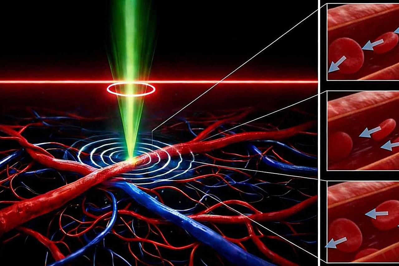
A map of DNA methylation changes in human organs could help researchers to discover more targets for anti-ageing therapies.
Researchers at Tsukuba University in Japan report that memories acquired while awake are stored in a more permanent form (called memory consolidation) during the REM stage of sleep, and that this process requires the reactivation of only a few specialized neurons involved in memory formation. They found that three of these neurons are crucial for memory consolidation during REM sleep.
The researchers focused on adult-born neurons (ABNs) in the hippocampal region of the temporal lobe, which are rare neurons known to be essential for maintaining proper memory function as the loss of these cells is observed in Alzheimer’s disease. However, it has remained unclear why the loss of this small neuronal population has such devastating effects on memory.
In the Nature Communications study, specially genetically modified mice, in which the activity of ABNs could be monitored, were exposed to a fear experience, and the researchers examined if the activities of these ABNs during initial memory formation were reproduced during REM sleep, when dreaming is believed to occur.
Researchers at the University of Illinois Urbana-Champaign have developed a novel framework for understanding and controlling the flow behavior of granular hydrogels—a class of material made up of densely packed, microscopic gel particles with promising applications in medicine, 3D bioprinting, and tissue repair.
The new study, published in Advanced Materials, was led by chemical and biomolecular engineering professors Brendan A. Harley and Simon A. Rogers, whose research groups specialize in biomaterials engineering and rheology, respectively.
Granular hydrogels have a unique ability to mimic the mechanical properties of living tissue, which makes them ideal candidates for encapsulating and delivering cells directly into the body. By integrating material synthesis and characterization with rheological modeling, the researchers created a predictive model that captures the essential physics of how granular hydrogels deform—reducing a complex problem to a few controllable parameters.
Scientists at Mount Sinai have created an artificial intelligence system that can predict how likely rare genetic mutations are to actually cause disease. By combining machine learning with millions of electronic health records and routine lab tests like cholesterol or kidney function, the system produces “ML penetrance” scores that place genetic risk on a spectrum rather than a simple yes/no. Some variants once thought dangerous showed little real-world impact, while others previously labeled uncertain revealed strong disease links.
A study published in Nature Ecology & Evolution reveals a surprising evolutionary insight: sometimes, losing genes rather than gaining them can help bacterial pathogens survive and thrive.
The study was conducted by a group of scientists and coordinated by Jaime Martínez Urtaza, from the Department of Genetics and Microbiology of the Universitat Autònoma de Barcelona (UAB); Yang Chao and Falush Daniel, from the Shanghai Institute of Immunity and Infection, Chinese Academy of Science; and Wang Hui, from the Shanghai Jiao Tong University.
When we think of evolution, we often imagine organisms changing or gaining new genes to adapt, such as growing wings, developing resistance, or evolving new behaviors. Across the tree of life, both spontaneous mutations and gene acquisition are classic tools of adaptation. However, in this study, researchers went down a lesser known and scarcely explored evolutionary path, the one of gene loss.
When genetic testing reveals a rare DNA mutation, doctors and patients are frequently left in the dark about what it actually means. Now, researchers have developed a powerful new way to determine whether a patient with a mutation is likely to actually develop disease, a concept known in genetics as penetrance.
The team set out to solve this problem using artificial intelligence (AI) and routine lab tests like cholesterol, blood counts, and kidney function. Details of the findings were reported in the journal Science. Their new method combines machine learning with electronic health records to offer a more accurate, data-driven view of genetic risk.
Traditional genetic studies often rely on a simple yes/no diagnosis to classify patients. But many diseases, like high blood pressure, diabetes, or cancer, don’t fit neatly into binary categories. The researchers trained AI models to quantify disease on a spectrum, offering more nuanced insight into how disease risk plays out in real life.
Using more than 1 million electronic health records, the researchers built AI models for 10 common diseases. They then applied these models to people known to have rare genetic variants, generating a score between 0 and 1 that reflects the likelihood of developing the disease.
A higher score, closer to 1, suggests a variant may be more likely to contribute to disease, while a lower score indicates minimal or no risk. The team calculated “ML penetrance” scores for more than 1,600 genetic variants.
Some of the results were surprising, say the investigators. Variants previously labeled as “uncertain” showed clear disease signals, while others thought to cause disease had little effect in real-world data.
Despite the association of regular coffee consumption with fewer neurodegenerative diseases, it remains unclear how coffee is associated with pre-clinical brain pathologies such as lesions in the white matter, degeneration of the cortex, or alterations of the microstructural integrity. White matter hyperintensities (WMH) are hyperintense lesions on T2-weighted images and are associated with an increased risk for stroke and depression, cognitive deterioration, and gait disorders [13,14,15]. As a marker of cerebral small vessel disease (CSVD) and vascular brain damage, WMH can vary in the degree of expression, depending on the age and the presence of cardiovascular risk factors, e.g., smoking or hypertension [16,17,18]. Previous studies have reported diverging results on the association of consumed coffee with imaging markers of CSVD. They found either beneficial associations of coffee with lacunar infarcts [7], beneficial [19] or detrimental [20] associations with WMH volume, or no significant associations at all [21,22].
A recently developed and valid imaging marker of microstructural integrity is the peak width of skeletonized mean diffusivity (PSMD), calculated as the distribution of the mean diffusivity (MD) between the 5th and 95th percentile in the white matter skeleton [23]. Only one study analyzed the association of coffee consumption with microstructural integrity, as quantified by fractional anisotropy, with a higher coffee consumption being associated with higher integrity of the white matter microstructure [24].
Damage to the brain structure is not restricted to white matter, but also extents to the cortex, e.g., in the form of atrophy. Except for one study focusing on the quantification of cortical thickness in regions susceptible for Alzheimer’s Disease [22], the link between coffee consumption and cortical thickness was only indirectly examined by measuring total brain volume or grey matter volume, with incongruent results between studies [7, 21,25,26]. This study aimed at investigating whether coffee consumption is associated with multiple brain MRI markers of vascular brain damage and neurodegeneration, including WMH, PSMD, and cortical thickness in a large, population-based cohort.
Scientists from Trinity College Dublin have discovered that electrically stimulating macrophages—one of the immune systems key players—can reprogram them in such a way as to reduce inflammation and encourage faster, more effective healing in disease and injury.
This breakthrough uncovers a potentially powerful new therapeutic option, with further work ongoing to delineate the specifics.
Macrophages are a type of white blood cell with several high-profile roles in our immune system. They patrol around the body, surveying for bugs and viruses, as well as disposing of dead and damaged cells, and stimulating other immune cells —kicking them into gear when and where they are needed.