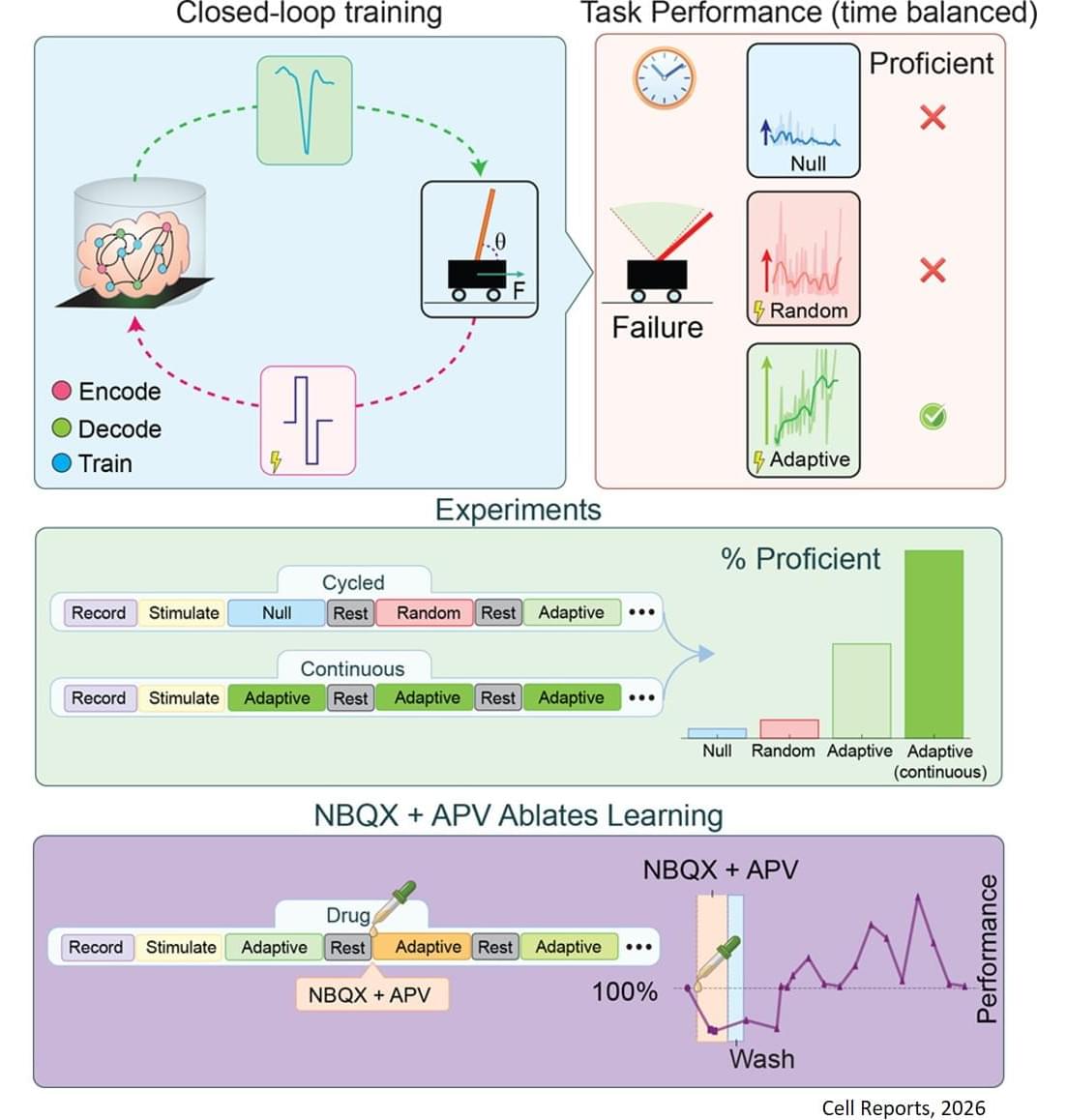
Toggle light / dark theme

How the nervous system activates repair after spinal cord injury

Through in vivo enhancer screening, the researchers also demonstrate that injury-responsive enhancers can selectively target reactive astrocytes across the CNS using therapeutically relevant gene delivery vectors.

“We have shown how cells read these instructions through a code that tells them how to react to injury. This code combines signals from general stress factors with the cell’s own identity,” explains the researcher.


After a spinal cord injury, cells in the brain and spinal cord change to cope with stress and repair tissue. A new study published in Nature Neuroscience, shows that this response is controlled by specific DNA sequences. This knowledge could help develop more targeted treatments.

When the central nervous system is damaged – for example, in a spinal cord injury – many cells become reactive. This means they change their function and activate genes that protect and repair tissue. However, how this process is regulated has long been unclear.

Researchers have now mapped thousands of so-called enhancers; small DNA sequences that act like ‘switches’ for genes, turning them on or boosting their activity.

World’s first 3D-printed brain tissue that mirrors human brain function

🧠💡 Thinking about organ transplants?

🔬 A team of scientists at the University of Wisconsin–Madison has achieved a groundbreaking milestone!

🌐 They’ve developed the world’s first 3D-printed brain tissue that mirrors human brain function.

🚀 This is a giant leap forward for research into neurological and neurodevelopmental disorders.

🖨️ Utilizing a horizontal layering technique and a softer bio-ink, this 3D-printing method allows neurons to weave together, forming networks similar to those in the human brain.

🔍 This precision in controlling cell types and arrangements opens new doors for studying neurological conditions, including Alzheimer’s and Parkinson’s disease.

Stem cell-mediated recovery in stroke: partnering with the immune system

In this Review, Saef Izzy and colleagues examine the therapeutic potential of stem cells in stroke, with a focus on neural and mesenchymal stem cells. They explore how these stem cells interact with brain immune cells to modulate the inflammatory microenvironment, restore blood–brain barrier integrity and promote tissue repair following a stroke.

Merging nanopores with nanofluidic devices could transform medicine and diagnostics

When disease begins forming inside the human body, something subtle happens long before symptoms appear. Individual molecules such as DNA, RNA, peptides, or proteins begin shifting in quantity or shape. Detecting these tiny molecular changes early could dramatically change how cancer, infections, and other conditions are diagnosed.

For years, scientists have dreamed of reading these changes one molecule at a time. Nanopores, nanometer-sized holes that detect single molecules by sensing changes in electric current, have brought that dream closer to reality, but nanopore sensing has hit several scientific walls.

Molecules rush through too fast to analyze. Signals drown in electrical noise. Proteins stick to pore surfaces and single pores simply cannot provide the scale required for real-world clinical use. So what would it take to make nanopore sensing fast, precise, and robust enough for society?

CRISPR Screens Revolutionize Human Neural Organoids Research

Studying the intricate molecular mechanisms that govern the assembly of the human nervous system has long been one of the most significant challenges in developmental biology and neuroscience. Researchers are continuously seeking a deeper understanding of how the human brain is built and what leads to various neurological disorders. Recent advancements in stem cell technology, particularly the ability to generate neural cells from pluripotent stem cells, coupled with the power of genome-editing tools like CRISPR-Cas9, are setting the stage for groundbreaking insights into human neurodevelopment and associated diseases. These technological innovations open new avenues for research that were previously thought to be unattainable.

The emergence of organoids and assembloids—miniature, simplified versions of brain tissue—has revolutionized the way scientists can model human development in vitro. Organoids replicate some of the complexity of human brain structures, allowing researchers to visualize developmental processes such as the specification, migration, and integration of neurons. This is particularly important for cortical interneurons, which migrate from the ventral forebrain to the dorsal forebrain during early brain development. These in vitro models provide an opportunity to study these intricate processes more closely and could lead to transformative discoveries in our understanding of brain diseases.

In a significant advancement outlined in recent research, scientists have developed a detailed protocol that marries pooled CRISPR-Cas9 screening with neural organoid and assembloid models. This innovative approach enables researchers to map hundreds of disease-related genes onto specific cellular pathways and critical aspects of human neural development. Such a strategy can significantly enhance our understanding of how various genes contribute to essential neuronal functions and the onset of neurological diseases, thereby paving the way for the development of novel therapeutic interventions.

Highly insulating polymer film that shields satellites to boost flexible electronics’ performance

Researchers have found that they could use highly insulating aluminum-coated polymer film to improve the performance of flexible electronics and medical sensors.

Currently, the aluminum-coated polymer film is used to shield satellites from temperature extremes.

Researchers at Empa have succeeded in making the material even more resistant by implementing an ultra-thin intermediate layer.

/* */