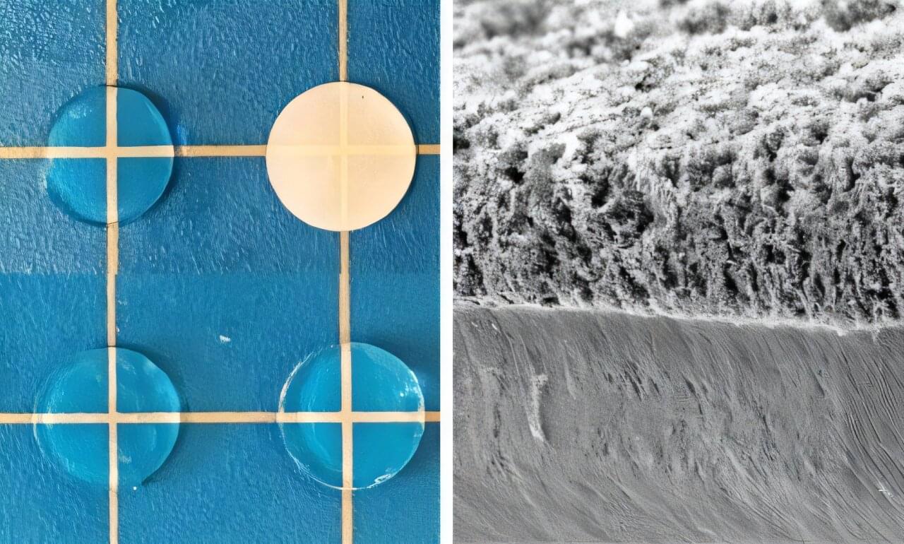
Toggle light / dark theme

Chemically ‘stapled’ peptides used to target difficult-to-treat cancers

Researchers at the University of Bath have developed a new technology that uses bacteria to build, chemically stabilize, and test millions of potential drug molecules inside living cells, making it much quicker and easier to discover new treatments for difficult-to-treat cancers.

Scientists based at the University’s Department of Life Sciences are investigating peptides—short chains of amino acids, the building blocks of proteins—as potential drugs for a family of notoriously “undruggable” cancer drivers known as transcription factors. These proteins act as master switches that control gene activity and are frequently overactive in cancer.

The Great Filter May Explain Why Civilizations Don’t Survive

The universe is old enough, large enough, and chemically rich enough to have produced countless civilizations. And yet, when we listen, we hear nothing. The Great Filter hypothesis offers one of the most disturbing explanations in modern science — somewhere between dead chemistry and starfaring intelligence, there exists a barrier so severe that almost nothing gets through. But the real question isn’t whether the filter exists. It’s whether we’ve already passed it — or whether it’s still ahead of us, waiting. This video explores the formal probability argument behind the silence, the candidate barriers hiding in the deep history of biology, the existential threats that scale with technological power, and what every new discovery about life beyond Earth actually tells us about our own survival odds.

Sources:
Robin Hanson, \

Read more

Why Ocean World Might Have Boiling Seas

“Not all of these satellites are known to have oceans, but we know that some do,” said Dr. Max Rudolph. [ https://www.labroots.com/trending/space/30266/ocean-world-boiling-seas-2](https://www.labroots.com/trending/space/30266/ocean-world-boiling-seas-2)


Could ocean worlds in the outer solar system have boiling water underneath their icy crusts? This is what a recent study published in Nature Astronomy hopes to address as a team of scientists investigated the geochemical processes that could be occurring on ocean worlds orbiting in the outer solar system. This study has the potential to help scientists better understand the conditions on ocean worlds throughout the solar system and where we can best search for life beyond Earth.

For the study, the researchers examined several icy moons orbiting Saturn and Uranus and what could happen as the ice shell on these moons becomes thinner over time. Specifically, they explored changes to the interior oceans beneath the icy shells, as some icy moons currently have oceans while others have evidence of past oceans that have since completely frozen over or escaped to space as water vapor.

In the end, the researchers identified different outcomes depending on the size of the moons. For example, if the ice shells on smaller moons like Saturn’s Mimas and Enceladus and Uranus’ Miranda become thinner, this could cause underlying oceans to boil from the decrease in pressure. However, if the ice shells on larger moons like Saturn’s Iapetus and Uranus’ Titania become thinner, this could lead to the ice shell collapsing, resulting in a type of plate tectonics.

New iron nanomaterial wipes out cancer cells without harming healthy tissue

Scientists at Oregon State University have engineered a powerful new nanomaterial that zeroes in on cancer cells and destroys them from the inside out. Designed to exploit cancer’s unique chemistry—its acidity and high hydrogen peroxide levels—the tiny iron-based structure sparks not one but two intense chemical reactions, flooding tumors with cell-damaging oxygen molecules. This dual attack overwhelms cancer cells with oxidative stress while sparing healthy tissue.

A universal spin–orbit-coupled Hamiltonian model for accelerated quantum material discovery

Zhong et al. introduce Uni-HamGNN, a graph neural network model that predicts spin–orbit-coupled electronic structures quickly and accurately, enabling fast screening and the discovery of advanced quantum materials across the periodic table.

Abstract: The changing landscape of urothelial carcinoma: on the edge of paradigm shift

In this Review Joshua J. Meeks discusses advancements in biomarkers and novel therapeutics that are likely to dramatically improve survival of patients with Bladder Cancer.


1Departments of Urology and.

2Biochemistry and Molecular Genetics, Northwestern University, Feinberg School of Medicine, Chicago, Illinois, USA.

3Jesse Brown VA Medical Center, Department of Veterans Affairs, Chicago, Illinois, USA.

Address correspondence to: Joshua J. Meeks, Department of Urology, Biochemistry and Molecular Genetics, Northwestern University, Feinberg School of Medicine, Chicago, Illinois, USA. Phone: 312.695.8146; Email: [email protected].

How Nanotech Made an Old Leukemia Drug 22,000x Stronger

Structural nanomedicine — what helped give us the COVID vaccine — may now be the key to a potent blood cancer treatment that’s had remarkable early results.


The findings, published in ACS Nano, show that just two doses of the experimental therapy achieved 97.5% tumor growth inhibition in a human AML xenograft mouse model — 59-fold more effective than standard 5-fluorouracil (5-FU) treatment, with no observable side effects.

For a disease with a grim 29% 5-year survival rate — and a cure rate of only 15% in patients older than 70 years — the findings offer a glimpse of how rethinking drug structure, not just chemistry, could advance cancer care.

Mirkin frames the findings within what he calls “the era of structural nanomedicine,” the idea that how you arrange medicinal components at the nanoscale matters as much as the molecules themselves.

Precision tumor imaging with a fluorescence probe and engineered enzymes

Successful cancer surgery depends on a surgeon’s ability to remove tumors, while minimizing harm to healthy tissues. Surgeons currently use glowing dyes which mark cancer cells to help differentiate from healthy cells, but these dyes aren’t perfect and will light up some healthy tissues too. For the first time, researchers including those from the University of Tokyo developed what they call a bioorthogonal fluorescence probe and a matching reporter enzyme that can activate the probe selectively at targeted tumor sites. This enables high-contrast tumor visualization with very low background. This study was performed in mice.

Cancer is a universal issue which affects uncountably many people around the world. Many will turn to surgery in the hope a surgeon will be able to completely remove a tumor leaving healthy tissues unaffected. Various tools and techniques have been developed over the years to improve the way these surgeries are performed, and visual imaging methods such as glowing dyes have proven to be very useful. But one drawback is that some probes can also be activated in healthy tissues by endogenous enzymes, creating background fluorescence and making it harder to judge what should be removed. The opposite is also possible, where cancer cells are left unmarked and are missed during surgery, increasing the chance of recurrence.

“Our group acknowledged this current shortcoming and improved upon this way to make cancer cells light up inside the body. In tests on mice, we delivered a special enzyme to tumors and used a fluorescence probe that only turns on when that enzyme is present,” said Associate Professor Ryosuke Kojima from the Laboratory of Chemical Biology and Molecular Imaging at the University of Tokyo. “Older probes often light up healthy tissue by mistake, creating background noise, but our highly selective, or bioorthogonal, dye probe is designed to stay completely off unless it meets its matching engineered enzyme. We essentially trained the enzyme through repeated mutation and selection, a form of directed evolution, so it could activate the probe strongly enough to work inside living animals.”

Smart fluorescent molecules provide cheaper path to sharper microscopy images

Multiphoton microscopy is used in biomedical research to study cells and tissues. Today, so-called two-photon microscopy is used to study processes within cells, but the technique has limitations in terms of image resolution. Four-photon microscopy provides images with higher resolution. However, such instruments are very expensive and, when studying biological material, the powerful laser light required can damage samples.

“In this project, we have developed molecules to visualize molecular-level details and monitor processes using the more common two-photon microscopy technique. These molecules have the capacity to achieve higher resolution than with four-photon microscopy, although two-photon microscopy is used,” says the project coordinator Joakim Andréasson, Professor at the Department of Chemistry and Chemical Engineering at Chalmers University of Technology.

“In the long term, results from studies of this kind may provide new insights into diseases, pharmaceuticals and the very smallest building blocks of life.”

A rewritable DNA hard drive may help solve the growing data storage crisis

Around the world, scientists are exploring an unexpected solution to the growing data crisis: storing digital information in synthetic DNA. The idea is simple but powerful—DNA is one of the most compact, durable information systems on Earth. But one issue has held the field back. Once data is written into DNA, it can’t be changed.

Now, researchers at the University of Missouri are helping to solve that problem by transforming DNA from a one-time medium into a rewritable digital hard drive. Their research is published in the journal PNAS Nexus.

“DNA is incredible—it stores life’s blueprint in a tiny, stable package,” said Li-Qun “Andrew” Gu, a professor of chemical and biomedical engineering at Mizzou’s College of Engineering. “We wanted to see if we could store and rewrite information at the molecular level faster, simpler and more efficiently than ever before.”

/* */