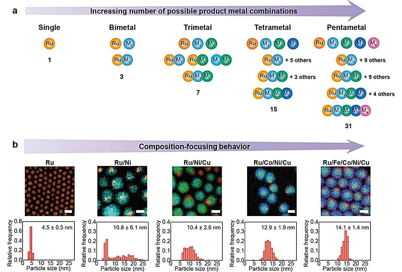
A nanocrystal is an extraordinarily tiny piece of material—composed of anywhere from a few to a few thousand atoms—in which atoms are arranged in a precise, ordered structure. Think of it like taking a piece of gold and shrinking it down to the size of a few hundred atoms. It’s still gold, still crystalline, just almost incomprehensibly small.
Nanocrystals are in the transistors inside computers and smartphones, in smartphone displays and TV screens, in the gold-nanoparticle sensors that power COVID and pregnancy tests, and in the pipes of your car exhaust system, among countless other innovations.
Their small size gives them a dramatically higher ratio of surface area to volume, making them especially useful as catalysts—materials that speed up chemical reactions without being consumed in the process.
Be kind to yourself this year. Using Zocdoc is FREE — visit my sponsor https://zocdoc.com/quinnsideas to find and instantly book an appointment with a top-rated, in-network doctor today.
Imagine a civilization reaches something like a Type II level, advanced enough to move through interstellar space and keep large populations alive for generations. At that stage, the challenge is developing ships that can cross the void, and also making sure the people inside them can survive radiation, isolation, and extreme travel times. That could mean heavy genetic engineering before the journey begins, changing bone density, metabolism, resistance to disease, tolerance for low gravity, or even sensory systems and respiration. But when they finally arrive, they may still find that the planet is wrong for them, maybe the air is toxic, the gravity is crushing, the temperatures are extreme, or the native chemistry is incompatible with human biology.
At that point, they face two paths. One is terraforming, which means trying to remake an entire planet into something closer to Earth. That could involve thickening or thinning an atmosphere, warming a frozen world, cooling a hot one, importing water, altering soil chemistry, introducing engineered microbes, building orbital mirrors or shades, and managing the planet for centuries or even millennia. The scale of that project is absurdly expensive, not just in money but in energy, infrastructure, labor, time, and raw materials. You are not changing a city or even a continent, you are trying to rewrite a whole world.
The other option is pantropy. Instead of forcing the planet to become Earth-like, the colonists change themselves to fit the planet. They might alter their lungs to breathe a different atmospheric mix, redesign their skin to handle harsher radiation, reduce their size for lower resource use, strengthen their bodies for higher gravity, or even become something so biologically different that they no longer look fully human. That is the core idea of pantropy, adapting the colonists to the world rather than adapting the world to the colonists.
The term was coined by James Blish, and he used it in connection with the stories collected in The Seedling Stars, especially “Surface Tension.” which was first published in 1952 in Galaxy Science Fiction.
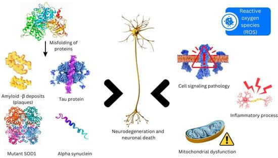
Neurodegenerative disorders entail a progressive loss of neurons in cerebral and peripheral tissues, coupled with the aggregation of proteins exhibiting altered physicochemical properties. Crucial to these conditions is the gradual degradation of the central nervous system, manifesting as impairments in mobility, aberrant behaviors, and cognitive deficits. Mechanisms such as proteotoxic stress, neuroinflammation, oxidative stress, and programmed cell death contribute to the ongoing dysfunction and demise of neurons. Presently, neurodegenerative diseases lack definitive cures, and available therapies primarily offer palliative relief. The integration of nanotechnology into medical practices has significantly augmented both treatment efficacy and diagnostic capabilities.
Researchers from the National University of Singapore (NUS) and collaborators have developed a predictive design strategy for creating graphene-like molecules with multiple interacting spins and enhanced resilience to magnetic perturbations, opening new avenues for molecular-scale quantum information technologies and next-generation spintronics.
The research team was led by Professor Lu Jiong from the NUS Department of Chemistry and the NUS Institute for Functional Intelligent Materials, together with Professor Wu Jishan from the NUS Department of Chemistry, and international collaborators, including key contributor Professor Pavel Jelínek from the Czech Academy of Sciences in Prague.
Magnetic nanographenes, which are molecules composed of fused benzene rings, are of growing interest for quantum technologies because they can host unpaired electrons, or spins, that may be used to store and process information. Unlike conventional magnetic materials based on metal atoms, these carbon-based systems offer chemical versatility and long spin coherence times. However, engineering a single molecule that contains multiple strongly coupled spins in a stable and controlled manner remains a major challenge.
Chemists and computer scientists tapped AI to find new disinfectants to combat the growing threat of dangerous “superbugs.”
The Journal of Chemical Information and Modeling published their computational-experimental framework for developing quaternary ammonium compounds, or QACs, to kill bacteria.
The method yielded 11 new QACs that show activity against antimicrobial-resistant bacteria.
Researchers at the University of Toronto have identified a protein from the quagga mussel that can stick to surfaces underwater, even though it lacks a chemical feature long thought to be essential for this kind of adhesion. The protein, called Dbfp7, is the first freshwater mussel adhesive protein to be functionally characterized.
The finding, published in PNAS, helps explain how some organisms attach themselves in wet environments and could inform the design of future medical glues—such as medical sealants and surgical adhesives—or other materials that need to work reliably in water.
Most studies of underwater adhesion have focused on marine mussels, which use proteins rich in a modified amino acid called 3,4-dihydroxyphenylalanine (DOPA) to bond to surfaces. Freshwater species have been studied less, and whether they rely on the same chemistry has not been clear.
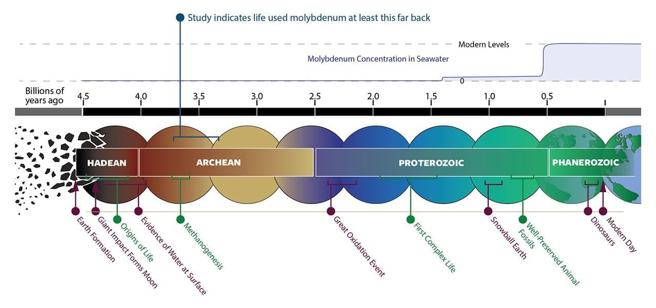
NASA-funded scientists have discovered that life on Earth over 3 billion years ago relied on the metal molybdenum, which was incredibly scarce in the environment at the time. The study, published in Nature Communications on Tuesday, is the first to show that molybdenum was used by ancient life this far back in our planet’s history.
On Earth today, molybdenum helps speed up vital biochemical reactions in cells. The metal is a component of essential enzymes that drive several major biological reactions in organisms. This is not only important for the individual organisms, but also biogeochemical cycles, such as the nitrogen cycle, which affect our entire planet. Without molybdenum, those important reactions could still happen in nature, but they would be too slow to sustain life.
“Molybdenum sits at the catalytic center of enzymes that run major carbon, nitrogen, and sulfur reactions,” explained Betül Kaçar, head of the Kaçar Lab at the University of Wisconsin-Madison and senior author on the study. Kaçar leads MUSE, a NASA Interdisciplinary Consortia for Astrobiology Research (ICAR) at UW-Madison.
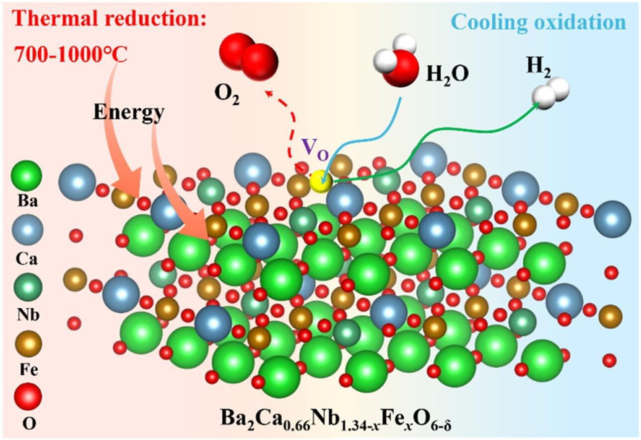
University of Birmingham research published today has shown a new low-temperature method for producing hydrogen that is suitable for both centralized hydrogen production, and also local generation using waste heat from large-scale industrial plants.
Hydrogen is the most abundant element in the universe and is a clean and environmentally friendly energy carrier. Unlike fossil fuels, which produce harmful emissions and carbon dioxide, it produces only heat and water on combustion and can also power fuel cells that produce electricity. But while hydrogen is carbon-free at the point of use, 95% of current production relies on fossil fuels.
Thermochemical splitting, where a catalyst splits water into hydrogen and oxygen, is emerging as a promising method for hydrogen production. However, current catalysts split water at 700‑1000oC and need temperatures between 1,300 and 1500oC to regenerate between cycles of water-splitting.
A collaborative team of scientists has discovered that life on Earth over three billion years ago relied on the metal molybdenum, which was incredibly scarce in the environment at the time. The study, published in Nature Communications, is the first to show that molybdenum was used by ancient life this far back in our planet’s history.
On Earth today, molybdenum helps speed up vital biochemical reactions in cells. The metal is a component of essential enzymes that drive several major biological reactions in organisms. This is not only important for individual organisms, but also biogeochemical cycles, such as the nitrogen cycle, which affect our entire planet. Without molybdenum, those important reactions could still happen in nature, but they would be too slow to sustain life.
“Molybdenum sits at the catalytic center of enzymes that run major carbon, nitrogen, and sulfur reactions,” explained Betül Kaçar, head of the Kaçar Lab at the University of Wisconsin-Madison and senior author on the study. Kaçar leads MUSE, a NASA Interdisciplinary Consortia for Astrobiology Research (ICAR) at UW-Madison.
Finding and developing new molecules is one of the great research endeavors of modern chemistry. From the development of new drugs to the creation of more sustainable materials, everything depends on finding new combinations of atoms with useful properties. Now, a research team from the Universitat Rovira i Virgili (URV) has developed an artificial intelligence tool capable of generating millions of new molecules which, although still unknown to science, comply with the laws of chemistry and could therefore be realistic possibilities. The research results have been published in the journal Nature Machine Intelligence.
The system, called CoCoGraph, works in a similar way to generative artificial intelligence tools for text or images, such as ChatGPT or Dall-E. “These models create new content that looks very much like the real thing. Our algorithm does the same, but with molecules,” explains Roger Guimerà, an ICREA Research Professor in the Department of Chemical Engineering at the URV.
Unlike other AI tools, however, the model does not yet respond to specific instructions. For the moment it simply carries out the more basic task of generating plausible molecules, that is, structures that comply with the rules of chemistry.