Start a career in Semiconductor Manufacturing with Merit America’s 14-week custom course with an industry-recognized certificate and $0 upfront. Learn more here!

Neuroendocrine cells are unique in their ability to act both as nerve cells and hormone-making cells. They’re scattered throughout the body, including the stomach, intestines, pancreas and lungs. Tumors that arise from these cells are called neuroendocrine tumors and are often rare and slow growing.
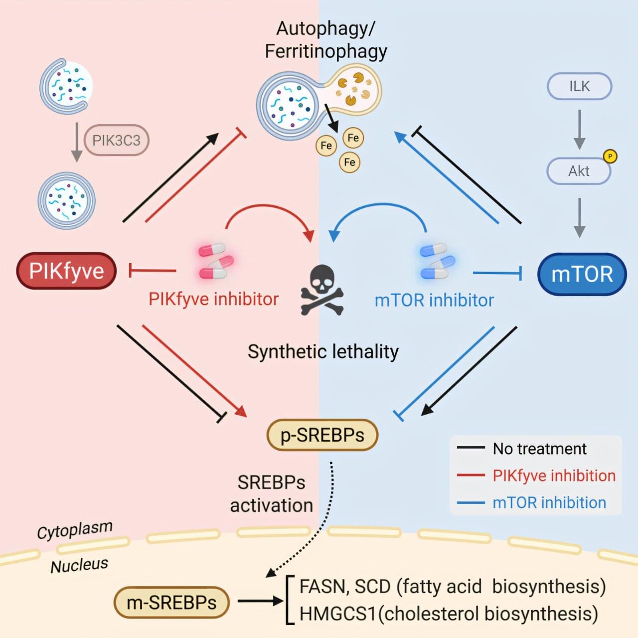
Around 70% of all neuroendocrine tumors arise in the pancreas or gastrointestinal tract and are known as gastroenteropancreatic neuroendocrine tumors, or GEP-NETs. Targeting these tumors is often challenging because cells become resistant to treatment.
In a recent study published in the journal Cell Reports Medicine, University of Michigan researchers have identified a new target that can suppress tumor growth. Their findings may lead to new treatment methods for GEP-NETs.
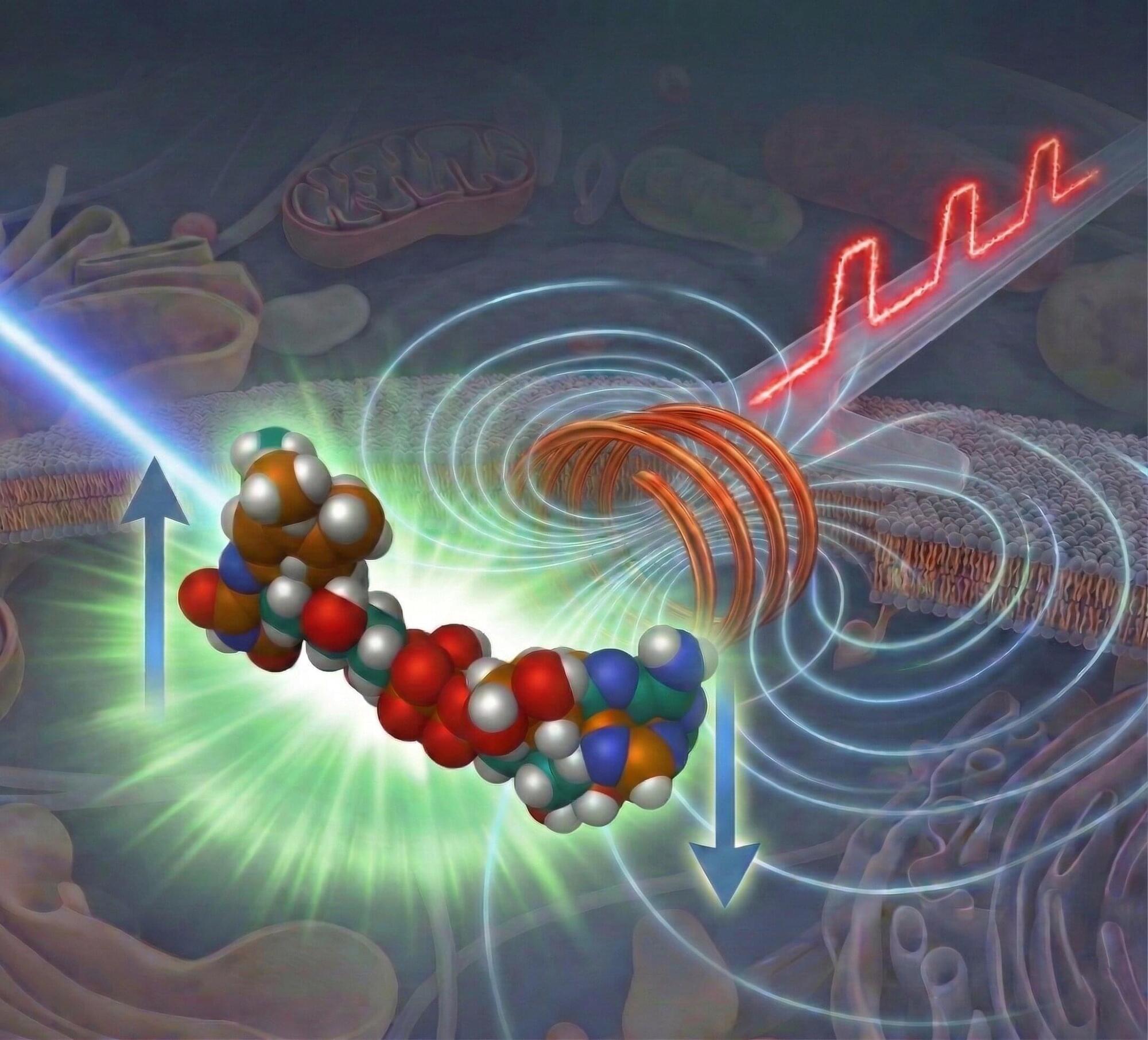
A research team at the University of Tokyo has developed a new microscopy platform that can observe a previously hidden layer of biomolecular chemistry linked to weak magnetic fields. The work, led by Project Researcher Noboru Ikeya and Professor Jonathan R. Woodward at the Graduate School of Arts and Sciences, addresses a long-standing technical gap in life-science measurement: Many important intermediates in spin-dependent reactions are “dark” molecules that do not emit light directly and therefore escape conventional fluorescence imaging.
To solve this, the team combined two precisely timed light pulses with a synchronized nanosecond magnetic pulse. The approach, called pump-field-probe fluorescence microscopy, compares signals as the magnetic field switches at different points in time. This comparison isolates the spin-dependent part of the chemistry and reveals precisely how magnetically sensitive intermediates appear and disappear. The findings are published in the Journal of the American Chemical Society.
The researchers validated the method in flavin-based model systems that are widely used to study biologically relevant photochemistry. They showed that the platform can recover reaction lifetimes and magnetic responses with high sensitivity, including at low concentrations matching cellular conditions. The system was capable of detecting very small signal changes under practical low-damage single-experiment per frame settings, an important step toward future live-cell studies.


Microsoft has resolved a known issue that was preventing some Classic Outlook users from sending emails via Outlook.com.
As the company explained when it acknowledged the issue last week, affected users were being warned that some of their messages hadn’t reached intended recipients.
Microsoft said that those experiencing this issue would encounter it more often when the Outlook.com account they used to send emails was an Outlook profile linked to another Exchange account.

Microsoft has deprecated and removed the Support and Recovery Assistant (SaRA) command-line utility from all in-support versions of Windows updates starting March 10.
SaRA is a free scriptable tool that helps troubleshoot and resolve common issues with Office, Microsoft 365, Outlook, and Windows by running a series of automated diagnostic tests on Windows 7, Windows 8, Windows 10, and Windows 11 systems.
According to Microsoft, the latest version of the utility should identify the root cause and then either automatically fix the issue, provide step-by-step instructions for a manual fix, or help users contact Microsoft support.