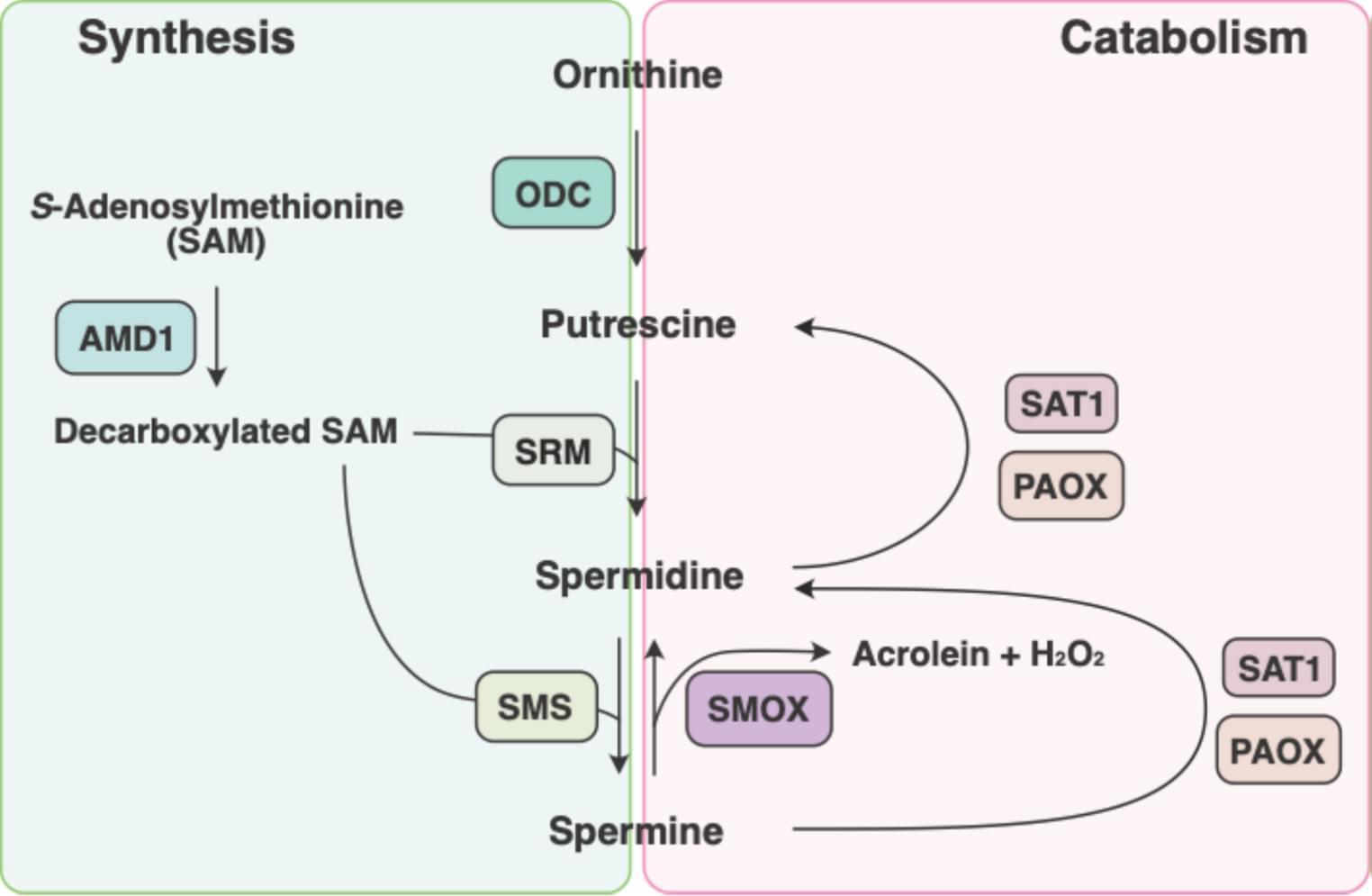
Toggle light / dark theme

A Machine‐Learning Approach Identifies Rejuvenating Interventions in the Human Brain

As the world population is ageing rapidly, with over two billion people projected to be above the age of 60 by 2050, age-related brain disorders are on the rise. Living longer but in poor health is not only a daunting prospect, it also places a substantial burden on healthcare systems worldwide. The idea of being able to counteract the functional decline of our brain through rejuvenating interventions sounds therefore promising. The question is how can we identify compounds that have the potential to efficiently rejuvenate brain cells and to protect the ageing population from neurodegeneration? Prof. Antonio Del Sol and his teams of computational biologists, based both at the LCSB from the University of Luxembourg and at the CIC bioGUNE in Bilbao, used their machine learning expertise to tackle the challenge.

The researchers developed what is called an “ageing clock”, a computational tool designed to measure the biological age of cells, as opposed to their chronological age. Indeed, the organs and tissues of people of the same age can evolve differently over time depending on genetic and environmental factors, leading to different biological ages. These clocks are therefore useful tools to assess ageing at the molecular level and can help in understanding its causes and consequences.

The clock designed by the LCSB and CIC bioGune researchers is specific to the brain and uses gene expression information from 365 genes to make predictions. Using a machine learning approach, it was trained on data from healthy individuals, aged from 20 to 97, and could accurately predict their age. Further tests showed that the clock is able to estimate the biological age of different cell types in the brain, especially neurons. Lastly, by looking at the predicted biological ages for healthy individuals and for patients with neurological conditions, the researchers observed that patients exhibited a higher biological age.

“Our results tell us that the biological age of the brain cells calculated by our clock reflects the decline in brain function experienced by the patients, especially between 60 and 70, and is even correlated with the degree of neurodegeneration,” explains Dr Guillem Santamaria, first author of the study. “It supports the view of neurodegeneration as a form of accelerated ageing but, more importantly, the positive association between neurodegeneration and biological age suggests that the rejuvenating interventions identified by the clock could serve as neuroprotective agents.”

The aim of the researchers was to use the clock to find genetic or chemical interventions that would significantly shift back the biological age of brain cells. They explored the effect of thousands of compounds on neural progenitor cells and neurons and identified 453 unique rejuvenating interventions.

Among the identified compounds that have the potential to reverse the biological age of the two types of brain cells, several are known to extend lifespan in animal models and some are already used to treat neurological disorders, but the vast majority has not yet been studied in the context of health-or lifespan extension. “On the one hand, the fact that our computational platform identified drugs that have a known effect on brain function supports the idea that using the predicted effect of a compound on the biological age is an efficient way to evaluate its neuroprotective potential,” details Prof. Antonio Del Sol, head of the Computational Biology groups at the LCSB and CIC BioGUNE. “On the other, the results also highlight that our clock can help us find many new candidates that haven’t been studied before for their rejuvenating properties. It opens up a lot of new avenues.”

As a proof of concept of their approach, the researchers then tested three of the predicted compounds in mice, in collaboration with the team of Prof. Rubén Nogueiras at the Centre for Research in Molecular Medicine and Chronic Diseases. The administration of these drugs significantly reduced anxiety and slightly increased spatial memory in older mice, addressing two well-known symptoms associated with ageing. An analysis of gene expression showed that the combination of these compounds also led to a shift toward a younger phenotype. Altogether, these results show that a selection of compounds predicted to rejuvenate the brain did produce rejuvenation at the molecular level in the cortex of aged mice and had an impact on behavioural and cognitive functions.

Globally, the study, recently published in the journal Advanced Science, highlights the computational ageing clock developed by the researchers as a valuable resource for identifying brain-rejuvenating interventions with therapeutic potential in neurodegenerative diseases. It provides a strong foundation for further research. “The hundreds of compounds predicted by our platform require validation across multiple biological systems to assess their efficacy and safety, offering extensive opportunities for future therapeutic development,” concludes Prof. Antonio Del Sol.

Genetic variants linked with higher risk of developing bipolar disorder

Bipolar disorder is a mental health condition characterized by extreme mood swings, with alternating periods of depression and manic episodes. Past research suggests that bipolar disorder has a strong genetic component and is among the most heritable psychiatric disorders.

To better understand the that increase the risk of developing this mental health disorder, neuroscientists and geneticists have carried out various genome-wide association studies (GWAS). These are essentially studies aimed at identifying specific regions of the human genome that are linked with an increased risk of having bipolar disorder, also referred to as bipolar risk loci.

While earlier works have identified many of these regions, causal single nucleotide polymorphisms (SNPs) for the disorder are largely unknown. These are essentially genetic variants that primarily contribute to bipolar disorder risk, as opposed to just being mere markers of it.

Can cancer drugs cure Alzheimer’s?

The team took publicly available data from three studies of the Alzheimer’s brain that measured single-cell gene expression in brain cells from deceased donors with or without Alzheimer’s disease. They used this data to produce gene expression signatures for Alzheimer’s disease in neurons and glia.

The researchers compared these signatures with those found in the Connectivity Map, a database of results from testing the effects of thousands of drugs on gene expression in human cells.

Out of 1,300 drugs, 86 reversed the Alzheimer’s disease gene expression signature in one cell type, and 25 reversed the signature in several cell types in the brain. But just 10 had already been approved by the FDA for use in humans.

Poring through records housed in the UC Health Data Warehouse, which includes anonymized health information on 1.4 million people over the age of 65, the group found that several of these drugs seemed to have reduced the risk of developing Alzheimer’s disease over time.

“Thanks to all these existing data sources, we went from 1,300 drugs, to 86, to 10, to just 5,” said the lead author of the paper.

The authors chose 2 cancer drugs out of the top 5 drug candidates for laboratory testing. They predicted one drug, letrozole, would remedy Alzheimer’s in neurons; and another, irinotecan, would help glia. Letrozole is usually used to treat breast cancer; irinotecan is usually used to treat colon and lung cancer.

The team used a mouse model of aggressive Alzheimer’s disease with multiple disease-related mutations. As the mice aged, symptoms resembling Alzheimer’s emerged, and they were treated with one or both drugs.

Decomposition of phenotypic heterogeneity in autism reveals underlying genetic programs

Classes of autism are uncovered with a generative mixture modeling approach leveraging matched phenotypic and genetic data from a large cohort, revealing different genetic programs underlying their phenotypic and clinical traits.

Study finds genetics shape health impact of leisure versus work physical activity

The benefits of exercise and its positive influence on physical and mental health are well documented, but a new Yale and VA Connecticut study sheds light on the role genetics plays for physical activity, accounting for some of the differences between individuals and showing differences in biology for physical activity at leisure versus physical activity at work and at home.

Using data from the Million Veteran Program (MVP), a genetic biobank run by the U.S. Department of Veterans Affairs, the researchers analyzed genetic influences on leisure, work, and home-time physical activity. They wanted to understand how genetics impacts these three types of physical activity and compare their health benefits.

The study included nearly 190,000 individuals of European ancestry, 27,044 of African ancestry, and 10,263 of Latin-American ancestry. To study the genetics of physical activity during leisure time, the researchers also added data from the UK Biobank, which included about 350,000 individuals.

Research uses AI to find pathologic and genetic basis for worse outcome of endometrial cancer in Black women

Endometrial cancer—in which tumors develop in the inner lining of the uterus—is the most prevalent gynecological cancer in American women, affecting more than 66,000 women a year. Black women are particularly at risk, with an 80% higher mortality rate than other demographic groups and a greater chance of contracting more aggressive cancer subtypes.

Regardless of lifestyle choices and health care equity, studies still show Black women have lower survival rates. A team of Emory researchers wondered: Could that poorer prognosis in Black women be caused by pathologic and genetic differences as well?

“Racism and equitable access to health care certainly play a big role in the increased mortality for populations of color,” says Anant Madabhushi, executive director of the Emory Empathetic AI For Health Institute. “But with endometrial cancer, it may not completely explain the difference in mortality.

Stem cell transplant without toxic preparation successfully treats genetic disease

An antibody treatment developed at Stanford Medicine successfully prepared patients for stem cell transplants without toxic busulfan chemotherapy or radiation, a Phase I clinical trial has shown.

While the researchers tested the protocol on patients with Fanconi anemia, a genetic disease that makes standard stem cell transplant extremely risky, they expect it may also work for patients with other genetic diseases that require stem cell transplants.

“We were able to treat these really fragile patients with a new, innovative regimen that allowed us to reduce the toxicity of the stem cell transplant protocol,” said the study’s co-senior author, Agnieszka Czechowicz, MD, Ph.D., assistant professor of pediatrics.

Finding Human Brain Genes in Duplicated DNA

“Historically, this has been a very challenging problem. People don’t know where to start,” said senior author Megan Dennis, associate director of genomics at the UC Davis Genome Center and associate professor in the Department of Biochemistry and Molecular Medicine and MIND Institute at the University of California, Davis.

In 2022, Dennis was a co-author on a paper describing the first sequence of a complete human genome, known as the ‘telomere to telomere’ reference genome. This reference genome includes the difficult regions that had been left out of the first draft published in 2001 and is now being used to make new discoveries.

Dennis and colleagues used the telomere-to-telomere human genome to identify duplicated genes. Then, they sorted those for genes that are: expressed in the brain; found in all humans, based on sequences from the 1,000 Genomes Project; and conserved, meaning that they did not show much variation among individuals.

They came out with about 250 candidate gene families. Of these, they picked some for further study in an animal model, the zebrafish. By both deleting genes and introducing human-duplicated genes into zebrafish, they showed that at least two of these genes might contribute to features of the human brain: one called GPR89B led to slightly bigger brain size, and another, FRMPD2B, led to altered synapse signaling.

“It’s pretty cool to think that you can use fish to test a human brain trait,” Dennis said.

The dataset in the Cell paper is intended to be a resource for the scientific community, Dennis said. It should make it easier to screen duplicated regions for mutations, for example related to language deficits or autism, that have been missed in previous genome-wide screening.

“It opens up new areas,” Dennis said.

New insights from the 1000 Genomes Project provide most complete view to date of human genetic variation

Completed in 2003, the Human Genome Project gave us the first sequence of the human genome, albeit based on DNA from a small handful of people. Building upon its success, the 1000 Genomes Project was conceived in 2007. The project began with the ambitious aim of sequencing 1,000 human genomes and exceeded it, publishing results gleaned from over 2,500 individuals of varying ancestries in 2015.

/* */