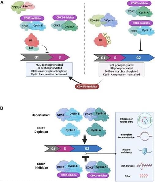
Toggle light / dark theme

Different genetic roots of autism may lead to shared brain activity and behaviors

New research from the University of Minnesota Medical School suggests that different genetic forms of autism may lead to similar patterns in brain activity and behavior. The findings were recently published in Nature Neuroscience.

Using brain-recording technology, the research team observed neurons across the entire brain to explore whether different genetic forms of autism share patterns and establish commonalities in neural responses. They found that, despite , various forms may show a similar unique pattern of —also known as a brain signature.

“We hope this research will serve as a stepping stone linking genetic differences and behavioral atypicalities,” said Jean-Paul Noel, Ph.D., an assistant professor at the University of Minnesota Medical School.

Aging on Chip: Harnessing the Potential of Microfluidic Technologies in Aging and Rejuvenation Research

Alternative models for studying aging have employed unicellular organisms such as the budding yeast Saccharomyces cerevisiae. Studying replicative aging in yeast has revealed insights into evolutionarily conserved enzymes and pathways regulating aging[ 12-14 ] as well as potential interventions for mitigating its effects.[ 15 ] However, traditional yeast lifespan analysis on agar plates and manual separation cannot track molecular markers and yeast biology differs from humans.[ 16 ]

Animal models, including nematodes, flies, and rodents, play a vital role in aging research due to their shorter lifespans and genetic manipulability, making them useful for mimicking human aging phenotypes.[ 17 ] These models have provided many insights into the fundamental understanding of aging mechanism. However, animal models come with several limitations when applied to human aging and age-related diseases. Key issues include limited generalizability due to species-specific differences in disease manifestation and physiological traits. For example, animal models often exhibit physiological differences, age at different rates, and may not fully replicate human conditions like cardiovascular disease,[ 18 ] immune response,[ 19 ] neurodegenerative diseases,[ 20 ] and drug metabolism.[ 21 ] Furthermore, in vivo models, such as rodents and non-human primates, suffer from limitations such as high costs, low throughput, ethical concerns, and physiological differences compared to humans. The use of shorter lifespan or accelerated aging models, along with the absence of long-term longitudinal data, can further distort the natural aging process and hinder our understanding of aging in humans. Additionally, many animal models rely on inbred strains, which lack genetic diversity and may not fully represent evolutionary complexity.[ 22 ]

In recent years, microfluidics has emerged as a promising tool for studying aging, offering of physiologically relevant 3D environments with high-throughput capabilities that surpass the limitations of traditional 2D cultures and bridge the gap between animal models and human As a multidisciplinary technology, microfluidics processes or manipulates small volumes of fluids (from pico to microliters) within channels measuring 10–1000 µm.[ 23 ] Traditional fabrication methods, such as photolithography and soft lithography, particularly using polydimethylsiloxane (PDMS), remain widely used due to their cost-effectiveness and biocompatibility. However, newer approaches, including 3D printing, injection molding, and laser micromachining, offer greater flexibility for rapid prototyping and the creation of complex architectures. Design considerations are equally critical and are tailored to the specific application, focusing on parameters such as channel geometry, fluid dynamics, material properties, and the integration of on-chip components like valves, sensors, and actuators. A comprehensive overview of the design and fabrication of microphysiological systems is beyond the scope of this review; readers are referred to existing reviews for further detail.[ 24-26 ] Microfluidic devices offer numerous advantages, including reduced resource consumption and costs, shorter culture times, and improved simulation of pathophysiological conditions in 3D cellular systems compared to other model systems (Figure 1).[ 27 ] Therefore, microfluidics platforms have been extensively employed in various domains of life science research, such as developmental biology, disease modeling, drug discovery, and clinical applications,[ 28 ] positioning this technology as a significant avenue in the field of aging research.

Scientists design a new tumor-targeting system for cancer fighting cells

CAR-T cells are specialized immune cells genetically modified to recognize and attack cancer cells. Researchers at Nagoya University in Japan and their collaborators have developed new CAR-T cells to target malignant tumors. While similar treatments have worked well for blood cancers, treating solid tumors is more difficult. Their method targeted a protein found in high amounts on many types of cancer cells (Eva1) and successfully eliminated tumors in lab mice.


A protein that appears on malignant tumors may hold the key to successful treatment.

De Novo Reconstruction of 3D Human Facial Images from DNA Sequence

Facial morphology is a distinctive biometric marker, offering invaluable insights into personal identity, especially in forensic science. In the context of high-throughput sequencing, the reconstruction of 3D human facial images from DNA is becoming a revolutionary approach for identifying individuals based on unknown biological specimens. Inspired by artificial intelligence techniques in text-to-image synthesis, it proposes Difface, a multi-modality model designed to reconstruct 3D facial images only from DNA. Specifically, Difface first utilizes a transformer and a spiral convolution network to map high-dimensional Single Nucleotide Polymorphisms and 3D facial images to the same low-dimensional features, respectively, while establishing the association between both modalities in the latent features in a contrastive manner; and then incorporates a diffusion model to reconstruct facial structures from the characteristics of SNPs. Applying Difface to the Han Chinese database with 9,674 paired SNP phenotypes and 3D facial images demonstrates excellent performance in DNA-to-3D image alignment and reconstruction and characterizes the individual genomics. Also, including phenotype information in Difface further improves the quality of 3D reconstruction, i.e. Difface can generate 3D facial images of individuals solely from their DNA data, projecting their appearance at various future ages. This work represents pioneer research in de novo generating human facial images from individual genomics information.

(Repost)


This study has introduced Difface, a de novo multi-modality model to reconstruct 3D facial images from DNA with remarkable precision, by a generative diffusion process and a contrastive learning scheme. Through comprehensive analysis and SNP-FACE matching tasks, Difface demonstrated superior performance in generating accurate facial reconstructions from genetic data. In particularly, Difface could generate/predict 3D facial images of individuals solely from their DNA data at various future ages. Notably, the model’s integration of transformer networks with spiral convolution and diffusion networks has set a new benchmark in the fidelity of generated images to their real images, as evidenced by its outstanding accuracy in critical facial landmarks and diverse facial feature reproduction.

Difface’s novel approach, combining advanced neural network architectures, significantly outperforms existing models in genetic-to-phenotypic facial reconstruction. This superiority is attributed to its unique contrastive learning method of aligning high-dimensional SNP data with 3D facial point clouds in a unified low-dimensional feature space, a process further enhanced by adopting diffusion networks for phenotypic characteristic generation. Such advancements contribute to the model’s exceptional precision and ability to capture the subtle genetic variations influencing facial morphology, a feat less pronounced in previous methodologies.

Despite Difface’s demonstrated strengths, there remain directions for improvement. Addressing these limitations will require a focused effort to increase the model’s robustness and adaptability to diverse datasets. Future research should aim to incorporating variables like age and BMI would allow Difface to simulate age-related changes, enabling the generation of facial images at different life stages an application that holds significant potential in both forensic science and medical diagnostics. Similarly, BMI could help the model account for variations in body composition, improving its ability to generate accurate facial reconstructions across a range of body types.

Genome of a 28-eyed jellyfish could provide insight on evolution of vision

One of the biggest mysteries of evolution is how species first developed complex vision. Jellyfish are helping scientists solve this puzzle, as the group has independently evolved eyes at least nine separate times. Different species of jellyfish have strikingly different types of vision, from simple eyespots that detect light intensity to sophisticated lens eyes similar to those in humans.

Biologists have studied jellyfish eye structure, light sensitivity, and visual behavior for decades, but the exact genes involved in jellyfish eye formation remain unknown.

Aide Macias-Muñoz, a professor of ecology and , is exploring how eyes and light detection evolved using genetic tools. Her lab has just completed a high-quality genome sequence of Bougainvillia cf. muscus, a small jellyfish-like animal in the Hydrozoa group that has an astonishing 28 eyes.

Is the Cell’s Antenna Related to Cancer Growth?

Many different types of cells in the body have a tiny projection known as a primary cilium. These cilia act like little signaling hub that can capture information about a cell’s environment and relay it to the cell, ultimately coordinating some cellular responses. The functions of cilia are well known in a few cases, such as in development, where they are crucial to the regulation of certain processes; or in some disorders called ciliopathies, in which genetic mutations lead to ciliary dysfunction and human disease.

Discovery of two new genetic disorders improves diagnoses for patients with neurodevelopmental conditions

The discovery of two new genetic disorders comes from a study delivered through the National Institute for Health and Care Research (NIHR) Manchester Biomedical Research Center (BRC) and The University of Manchester and could provide answers for several thousands of people with neurodevelopmental conditions around the world.

Since the breakthrough, 18-year-old Rose Anderson from Stretford in Manchester has received a diagnosis of one of the newly discovered conditions.

Rose has been known to the team at the Manchester Center for Genomic Medicine at Manchester University NHS Foundation Trust (MFT) for nearly her whole life, although a precise diagnosis for her seizures and has proved difficult to find.

/* */