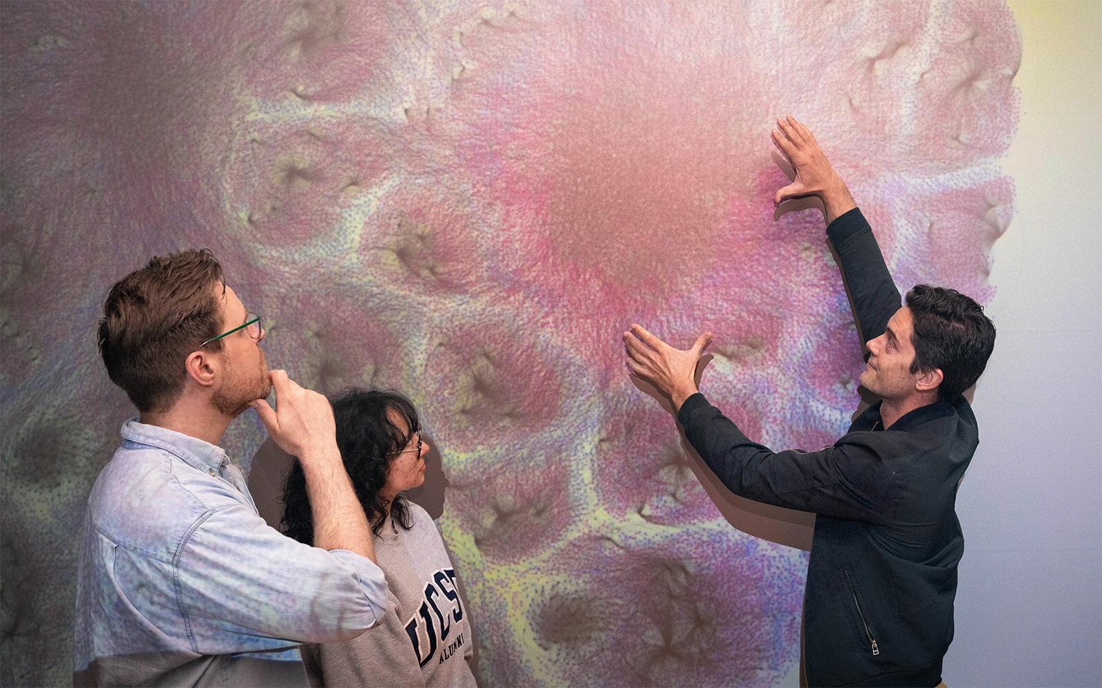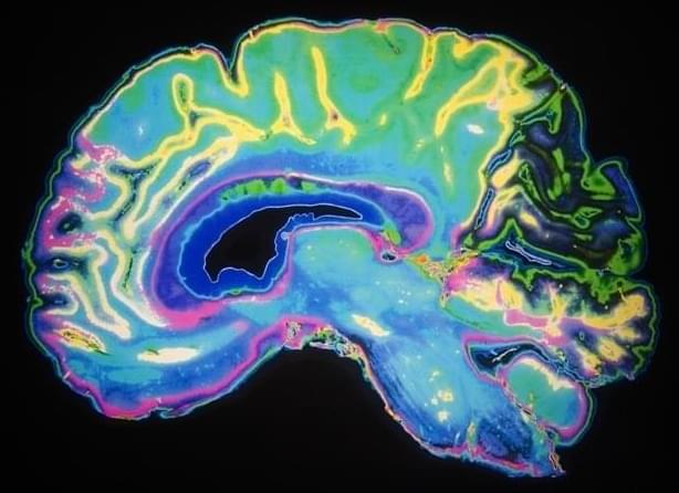
Johns Hopkins scientists say they have used 3D imaging, special microscopes and artificial intelligence (AI) programs to construct new maps of mouse brains showing a precise location of more than 10 million cells called oligodendrocytes. These cells form myelin, a protective sleeve around nerve cell axons, which speeds transmission of electrical signals and support brain health.
Published online Feb. 18 in Cell and funded by the National Institutes of Health, the maps not only paint a whole-brain picture of how myelin content varies between brain circuits, but also provide insights into how the loss of such cells impacts human diseases such as multiple sclerosis, Alzheimer’s disease and other disorders that affect learning, memory, sensory ability and movement, say the researchers. Although mouse and human brains are not the same, they share many characteristics and most biological processes.
“Our study identifies not only the location of oligodendrocytes in the brain, but also integrates information about gene expression and the structural features of neurons,” says Dwight Bergles, Ph.D., the Diana Sylvestre and Charles Homcy Professor in the Department of Neuroscience at the Johns Hopkins University School of Medicine. “It’s like mapping the location of all the trees in a forest, but also adding information about soil quality, weather and geology to understand the forest ecosystem.”