If you want to own a piece of a longevity technology company, here is your chance.
Invest as little as $100 in startups and small businesses. Wefunder is the largest Regulation Crowdfunding portal.

In a research paper published in Nature Aging, the team reports using a novel approach to provide the first data-driven classification of multiple diseases obtained using human genetic and medical data freely available from the UK Biobank.
Co-author Professor Linda Partridge (UCL Institute of Health Aging and Max Planck Institute for Biology of Aging) said: Advancing age is the main risk for major diseases, including cancer, dementia, and cardiovascular disease. Understanding the molecular links between the aging process and age-related diseases could allow them to be targeted with drugs to improve late-life health.
The striking finding from the study was that diseases with a similar age of onset were genetically more similar to each other than they were to diseases in the other three clusters.
Papers referenced in the video:
Dietary Thiols: A Potential Supporting Strategy against Oxidative Stress in Heart Failure and Muscular Damage during Sports Activity:
https://www.ncbi.nlm.nih.gov/pmc/articles/PMC7765667/
Ergothioneine levels in an elderly population decrease with age and incidence of cognitive decline; a risk factor for neurodegeneration?
https://pubmed.ncbi.nlm.nih.gov/27444382/
Is ergothioneine a ‘longevity vitamin’ limited in the American diet?
https://www.ncbi.nlm.nih.gov/pmc/articles/PMC7681161/
Frailty markers comprise blood metabolites involved in antioxidation, cognition, and mobility:
https://pubmed.ncbi.nlm.nih.gov/32295884/
Ergothioneine is associated with reduced mortality and decreased risk of cardiovascular disease:

Scientists injected dozens of human stem cells into developing monkey embryos, and the resulting hybrids survived for up to 20 days in lab dishes.
These human-monkey embryos could someday serve as helpful models for human disease, embryonic development and aging, the study authors noted in a new report, published April 15 in the journal Cell. By zooming in on the interaction of human and animal cells in the embryos, scientists could also learn how to help human cells survive amongst animal cells, potentially advancing the effort to grow human organs in living animal models.
The waterless toilet was developed by Cranfield University.
The toilet was developed for use in countries that doesn’t have running water.
This toilet doesn’t smell which is a result of multiple actions that go on beneath the toilet once the lid is closed.
–A set of gears within the toilet are turned when the lid is closed, these gears rotate the basin where the fecal delight was deposited.
–The waste falls into a holding chamber where a swipe blade wipes the inside of the holding basin.
–The solids drop down to the bottom, and the liquid floats to the top.
–An archimedes screw carries the waste upwards where it gets rolled into pellets that drop into a combustor and are burned.
–The combustor on the toilet will be on all the time but will require an initial source of power to get it going. The team had an initial idea of attaching a hand crank or a bicycle to generate the power needed but recently scrapped that idea. A solar panel could be installed above the toilet but that wouldn’t be very cost effective. The team has other ideas they are still working through to solve this issue.
–The poop ash that is accumulated from the combustor needs to be removed once a week.
–The liquid floats through a set of pipes that are above the combustor.
–The liquid is heated and passed through a set of 4 membrane bundles that purifies the water.
–This purified water drips down to the bottom where it travels to and is stored in the front step of the toilet.
–This water while purified isn’t clean enough to drink but it can be used in the garden to grow plants as well as for cleaning.
–This waterless toilet needs to be serviced every 3 months, the 4 membranes need to be replaced to continue to purify the water.
–So now you have the full overview of this toilet what problems do you guys foresee with this invention?
–The only thing I’m going to say is that I’m sure the swipe blade will wipe most of the poop out of the holding basin, you know there’s gonna be some nasty skip marks in there.
In reality though places in the world where people have real struggles in life, a skip mark isn’t one of them.
Sources:
https://www.youtube.com/watch?v=jGPpXF7y9Rg.
http://www.sciencedirect.com/science/article/pii/S0196890416306628
Check out some of our other videos:
Top 10 Fruits You’ve Never Heard Of Part 2
https://www.youtube.com/watch?v=hRdgPyZF45g&feature=youtu.be.
Top 10 Fruits You’ve Never Heard Of.
https://www.youtube.com/watch?v=OKTej1u-7-0&feature=youtu.be.
Top 5 Most EXTREME Roller Coasters.
https://www.youtube.com/watch?v=sTofF4_OPkM&feature=youtu.be.
Top 10 Craziest Lego Creations – Lego Sculptures to Blow Your Mind!
https://www.youtube.com/watch?v=mip9G0_rUTk&feature=youtu.be.
Top 10 LONGEST LIVING Creatures on Earth.
https://www.youtube.com/watch?v=IDaPH_ZGiVw&feature=youtu.be.
10 SHOCKING Photos That Will Leave You SPEECHLESS
https://www.youtube.com/watch?v=k1PdOc8QjR4&feature=youtu.be.
Top 10 DIRTIEST Public Surfaces you TOUCH!
https://www.youtube.com/watch?v=d0j8CQTXkF0&feature=youtu.be.
Support TTL by using our amazon link: http://amzn.to/2dQQ4nu.
Check us out on social media:
Donna butts — executive director, generations united — focusing on the psycho-social aspects of healthy aging and wellness.
Donna Butts is the Executive Director of Generations United, an organization with a mission to improve the lives of children, youth, and older people through intergenerational collaboration, public policies, and programs for the enduring benefit of all, a position she has held since 1997. For more than 30 years, Ms. Butts has worked tirelessly to promote the well-being of children, youth and older adults through nonprofit organizations across the country and around the world. She began her career in her home state of Oregon as a youth worker with the YWCA, where she worked one-on-one with teens and saw the positive effects of intergenerational programs firsthand.
Ms. Butts has held leadership positions with Covenant House, a New York-based international youth serving organization, and the National 4-H Council. She served as the Executive Director for the National Organization on Adolescent Pregnancy, Parenting and Prevention before taking the helm of Generations United.
An internationally sought-after speaker, author and advocate, Ms. Butts frequently speaks on intergenerational connections, grandparents raising grandchildren and policies effective across the lifespan. Her commentary has appeared in the New York Times, the Washington Post, the Christian Science Monitor and the Wall Street Journal. She has been interviewed on the TODAY Show, National Public Radio and ABC News, and was invited by the United Nations to sit on four expert panels most recently on intergenerational solidarity and social cohesion in preparation for the 2014 20th anniversary of the International Year or the Family.
In 2004, Ms. Butts was honored with the National Council on Aging’s Jack Ossofsky for Leadership, Creativity, and Innovation in Programs and Services for Older Persons. She served as a 2005 delegate to the White House Conference on Aging. A respected author, she has written countless articles, chapters and publications regarding the welfare of children, youth and older adults.
How seriously do you take your diet?
It is one of the foundations upon which everything else stands.
We have gone from abundant quality in a limited supply in our evolutionary past, to an abundance of quantity but with the quality now lacking (for many), and yet, as I show here, your health, future prospects for disease and illness, and ultimately, your longevity, are intrinsically linked.
In this video I take it right back to ground zero, and then bring the latest studies to bear, so we can see how it influences it all, so you can make the right decisions about what you should, and maybe should not, be eating.
Hope you find it of interest.
Have an amazing day!!

Michael D. West talking about regeneration.
[Reuploaded from Vimeo: https://vimeo.com/522148118]
In this video, Dr. Michael West, CEO of AgeX Therapeutics, discusses the non-peer reviewed preprint article in bioRxiv on the potential role of the clustered protocadherin genes in regeneration, aging, and cancer, and the relevance of the discovery for iTR product development.
Full paper: https://www.biorxiv.org/content/10.1101/2021.03.07.434314v1.full.
Aubrey de Grey’s talk during the South Summit that took place in Spain last October 2020. Aubrey explains why he thinks science and technology is close to bringing aging under complete medical control.
He also describes how along the process we will reach what he calls “Longevity Escape Velocity”. Once we reach it, we will be able to stay one step ahead of the curve of aging, and extend significantly, eventually indefinetely, human health and lifespan.
During the South Summit that took place in Spain last October 2020, gerontologist Aubrey de Grey explains why he thinks science and technology is close to bringing aging under complete medical control.
He also describes how along the process we will reach what he calls longevity escape velocity. Once we reach it, we will be able to stay one step ahead of the curve of aging, and extend significantly, eventually indefinetely, human health and lifespan.
El video cuenta con subtítulos en Español.
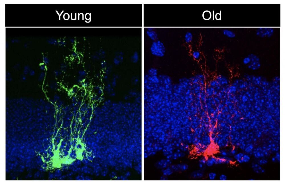
In a new study published in Cell Stem Cell, a team led by USC Stem Cell scientist Michael Bonaguidi, Ph.D., demonstrates that neural stem cells—the stem cells of the nervous system—age rapidly.
“There is chronological aging, and there is biological aging, and they are not the same thing,” said Bonaguidi, an Assistant Professor of Stem Cell Biology and Regenerative Medicine, Gerontology and Biomedical Engineering at the Keck School of Medicine of USC. “We’re interested in the biological aging of neural stem cells, which are particularly vulnerable to the ravages of time. This has implications for the normal cognitive decline that most of us experience as we grow older, as well as for dementia, Alzheimer’s disease, epilepsy and brain injury.”
In the study, first author Albina Ibrayeva, a Ph.D. candidate in the Bonaguidi Lab in the Eli and Edythe Broad Center for Regenerative Medicine and Stem Cell Research at USC, joined her colleagues in looking at the brains of young, middle-aged and old mice.