Toggle light / dark theme

Novel carbon nanotube-based transistors reach THz frequencies

Carbon nanotubes (CNTs), cylindrical nanostructures made of carbon atoms arranged in a hexagonal lattice, have proved to be promising for the fabrication of various electronic devices. In fact, these structures exhibit outstanding electrical conductivity and mechanical strength, both of which are highly favorable for the development of transistors (i.e., the devices that control the flow of current in electronics).

In recent years, several have started using CNTs to develop various electronics, including metal-oxide-semiconductor field-effect transistors (MOSFETs). MOSFETs are transistors that control the flow of current through a semiconducting channel utilizing an applied to a gate electrode.

Notably, when arrays of CNTs are used to develop MOSFETs, they can operate at (RF), the range of electromagnetic waves that support wireless communication. The resulting MOSFETs could thus be particularly advantageous for the advancement of wireless communication systems and technologies.

Development of revolutionizing photo-induced microscopy and its use around the globe celebrated in new publication

Photo-induced force microscopy began as a concept in the mind of Kumar Wickramasinghe when he was employed by IBM in the early years of the new millennium. After he came to the University of California, Irvine in 2006, the concept evolved into an invention that would revolutionize research by enabling scientists to study the fundamental characteristics of matter at nanoscale resolution.

Since the earliest experimental uses of PiFM around 2010, the device, which reveals the chemical composition and spatial organization of materials at the , has become a tool of choice for researchers in fields as diverse as biology, geology, materials science and even advanced electronics manufacturing.

“This is the story of a technology that was inspired by work at IBM, was invented and developed at UC Irvine, then got spun off, and now we have instruments on all continents across the world except for Antarctica,” says Wickramasinghe, Henry Samueli Endowed Chair and Distinguished Professor emeritus of electrical engineering and computer science who now holds the title of UC Irvine Distinguished Research Professor. “Almost anywhere serious research is happening, there are people out there who are using PiFM to discover new things.”

Nanoparticle blueprints reveal path to smarter medicines

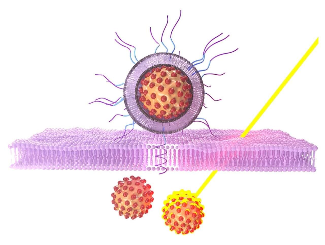
Lipid nanoparticles (LNPs) are the delivery vehicles of modern medicine, carrying cancer drugs, gene therapies and vaccines into cells. Until recently, many scientists assumed that all LNPs followed more or less the same blueprint, like a fleet of trucks built from the same design.

Now, in Nature Biotechnology, researchers from the University of Pennsylvania, Brookhaven National Laboratory and Waters Corporation have characterized the shape and structure of LNPs in unprecedented detail, revealing that the particles come in a surprising variety of configurations.

That variety isn’t just cosmetic: As the researchers found, a particle’s internal shape and structure correlates with how well it delivers therapeutic cargo to a particular destination.

Magnetized plasmas offer a new handle on nanomaterial design

Imagine a cloud that shines like a neon sign, but instead of raindrops, it contains countless microscopic dust grains floating in midair. This is a dusty plasma, a bizarre state of matter found both in deep space and in the laboratory.

In a new study, published this week in Physical Review E, Auburn University physicists report that even can reshape how these dusty plasmas behave—slowing down or speeding up the growth of nanoparticles suspended inside. Their experiments show that when a magnetic field nudges into spiraling paths, the entire plasma reorganizes, changing how particles charge and grow.

“Dusty plasmas are like in a vacuum box,” said Bhavesh Ramkorun, lead author of the study. “We found that by introducing magnetic fields, we could make these particles grow faster or slower, and the ended up with very different sizes and lifetimes.”

Exosomes as critical mediators of cell-to-cell communication in cancer pathogenesis and their potential clinical application

Extracellular vesicles can be divided into 3 primary classes based on their size: exosomes (20–100 nm), microvesicles (100–1,000 nm) and apoptotic bodies (1–5 µm). Exosomes have been the major focus of extracellular vesicle research. The term “exosome” was coined by Trams et al. in 1981 for “exfoliated membrane vesicles with 5’-nucleotidase activity” (23). Exosomes are distinguished from apoptotic bodies and microvesicles in term of their size, origin (endosomal or cell membrane), markers and composition. With spherical to cup-shaped nanoparticles and specific surface molecular markers, such as CD9 and CD63, exosomes are formed by the inward budding of endosomal membranes, thereby containing a variety of proteins, mRNAs and miRNAs (24-27). In addition to various cell or tissue specific materials, exosomes also contain certain common proteins, including cytoplasmic proteins (Hsp70 and Hsp90), cytoskeletal proteins (tubulin and actin), membrane fusion proteins (Rab GTPases) and membrane-associated proteins (CD9, CD81 and CD63) (28-30). These proteins could be used as markers for exosome isolation and identification. However, exclusive protein markers for exosomes are currently unknown. The material contained in exosomes is well protected to prevent degradation. For example, the RNA in exosomes is more stable than that in plasma and is not easily degraded by RNases. Exosomal RNA can be stored at −20 °C for more than 5 years, and the concentration is not decreased when compared with freshly prepared samples (31).

The signals and mechanisms underlying exosome formation and cargo sorting into exosomes have not been thoroughly elucidated to date. The present evidence shows that at least Endosomal Sorting Complexes Required for Transport (ESCRT) class proteins, tetraspanin CD63, specific glycan modification, the p53/TSAP6 pathway, and/or lipid-dependent mechanisms are involved in the formation of intraluminal vesicles in extracellular vesicles (32). Moreover, Rab-dependent trafficking mechanisms (Rab11, Rab27 and Rab35) have roles in exosome exocytosis and secretion (33) (Figure 1). Recipient cells internalize the foreign exosomes via multiple processes, including phagocytosis, clathrin-mediated endocytosis, macropinocytosis, and receptor-mediated and direct fusion (34,35). The factors that determine which and how a molecule is included or excluded in exosomes is under debate. It is reported that as a component of the COP9 signalosome regulatory complex, JAB1/CSN5 is involved in sorting proteins into exosomes (36). The introduction of exosomes provides a new molecular platform to further study cell-cell interaction, specific targeted cell selection, mechanisms of internalization and the potential of serving as a drug delivery system (37,38). Moreover, exosomes have been found in nearly all human body fluids, such as blood plasma, saliva, cerebrospinal fluid, urine, malignant ascites and semen (39-42), thereby implying that exosomes can be exploited as useful tools for cancer diagnosis and predictive biomarkers for cancer prognosis. It is interesting that the rate of exosomal release and content is different between healthy cell exosomes and tumor-derived exosomes. Numerous studies, including in vitro and in vivo studies, as well as clinical analysis, demonstrate that the number of exosomes increases significantly in cancer cells compared to normal cells. The distinct content of exosomes between the two groups (most notably miRNAs) may have important clinical significance (43,44).

Sensor identifies sodium nitrite in drinks using laser-modified cork

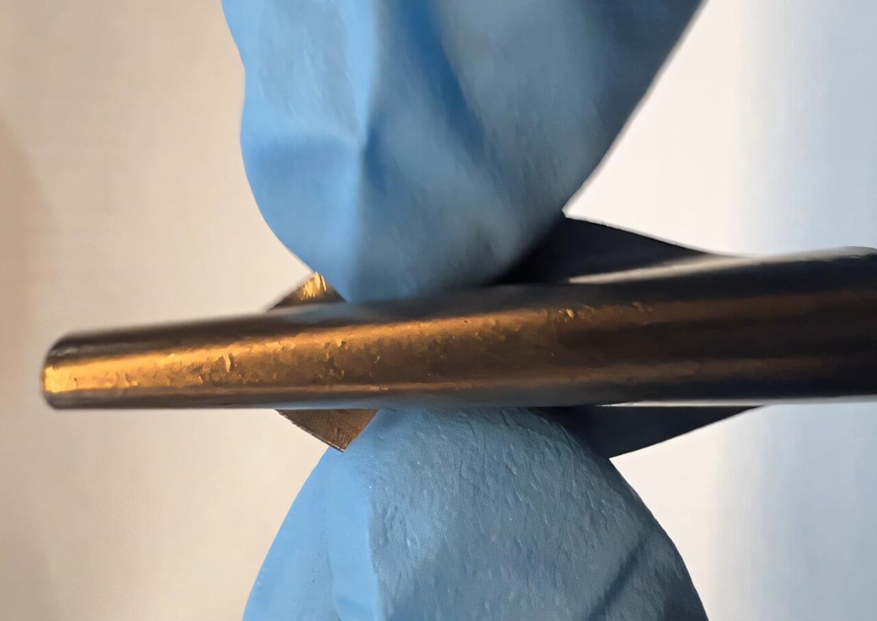
A team of researchers from the Federal University of São Carlos (UFSCar) in the state of São Paulo, Brazil, has developed a sensor that can identify sodium nitrite (NaNO2) in various beverages, including mineral water, orange juice, and wine. This inorganic salt is used as a preservative and fixative to give products such as ham, bacon, and sausages their pink or red color. Depending on the amount, it can cause serious health problems by leading to the formation of nitrosamines, which are carcinogenic compounds.

“This risk motivated us to develop a simple, fast, and accessible way to detect the compound and ensure the quality and safety of liquid consumption,” says Bruno Campos Janegitz, head of the Laboratory of Sensors, Nanomedicine, and Nanostructured Materials (LSNano) at UFSCar. Janegitz coordinated the study, which was published in the journal Microchimica Acta.

“Detection [of NaNO2] in beverages, especially wines, is important for , since its use is not legally permitted in Brazil and most countries,” the authors write in the article.

Ultrasensitive sensor maps magnetization textures in rhombohedral graphene

Graphene, which is comprised of a single layer of carbon atoms arranged in a hexagonal lattice, is a widely used material known for its advantageous electrical and mechanical properties. When graphene is stacked in a so-called rhombohedral (i.e., ABC) pattern, new electronic features are known to emerge, including a tunable band structure and a non-trivial topology.

Due to these emerging properties, electrons in rhombohedral can behave as if they are being influenced by “hidden” magnetic fields, even if no is applied to them. While this interesting effect is well-documented, closely studying how electrons organize themselves in the material, with their spins and valley states pointing in different directions, has so far proved challenging.

Researchers at Weizmann Institute of Science recently set out to further examine the local magnetization textures in rhombohedral graphene, using a nanoscale superconducting quantum interference device (nano-SQUID). Their paper, published in Nature Physics, offers new insight into the physical processes governing the correlated states previously observed in the material.

Curved nanosheets in anode help prevent battery capacity loss during fast charging

As electric vehicles (EVs) and smartphones increasingly demand rapid charging, concerns over shortened battery lifespan have grown. Addressing this challenge, a team of Korean researchers has developed a novel anode material that maintains high performance even with frequent fast charging.

A collaborative effort by Professor Seok Ju Kang in the School of Energy and Chemical Engineering at UNIST, Professor Sang Kyu Kwak of Korea University, and Dr. Seokhoon Ahn of the Korea Institute of Science and Technology (KIST) has resulted in a hybrid anode composed of graphite and organic nanomaterials. This innovative material effectively prevents capacity loss during repeated fast-charging cycles, promising longer-lasting batteries for various applications. The findings are published in Advanced Functional Materials.

During battery charging, lithium ions (Li-ions) move into the , storing energy as Li atoms. Under rapid charging conditions, excess Li can form so-called “dead lithium” deposits on the surface, which cannot be reused. This buildup reduces capacity and accelerates battery degradation.

/* */