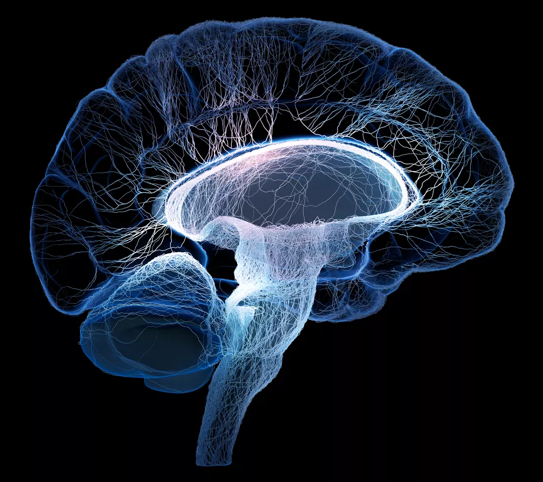
New research on brainwaves in the thalamus shows patterns distinctly linked to conscious states.

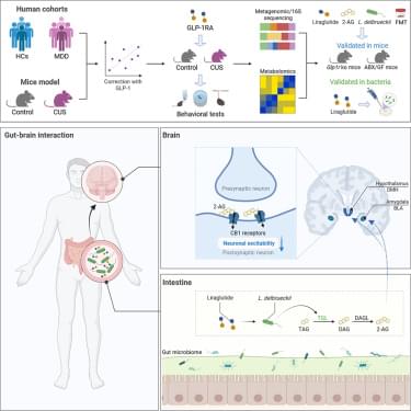
To determine whether canonical GLP-1R signaling is required for liraglutide to remodel the gut microbiota, we performed 16S rRNA sequencing on fecal samples from CUS-exposed wild-type (WT) and Glp1r−/− mice treated with or without liraglutide. Analyses of alpha-diversity, beta-diversity, and genus-level composition revealed that liraglutide changed the microbial structure in CUS mice, although specific compositional shifts differed between WT and Glp1r−/− mice (Figure S6). However, linear discriminant analysis (LDA) identified the genus Lactobacillus as the most significantly enriched taxon following liraglutide treatment in both WT and Glp1r−/− mice (Figures 2 H and 2I). Consistent with this finding, the abundance of Lactobacillus, which was reduced by CUS, was restored by liraglutide in both WT and Glp1r−/− mice (Figure 2 J). To identify the specific Lactobacillus species affected, we performed metagenomic sequencing on fecal samples from CUS mice treated with liraglutide. The Venn diagram showed that L. delbrueckii emerged as the most markedly altered species following liraglutide intervention in CUS mice (Figures 2 K and 2L). Targeted qPCR further validated that CUS-induced reduction in L. delbrueckii abundance was restored by liraglutide treatment in both WT and Glp1r−/− mice (Figures S7 A and S7B). Moreover, semaglutide, another GLP-1R agonist, similarly reversed the CUS-induced reduction of L. delbrueckii, suggesting a shared effect within this class of drugs (Figure S7 C). Together, these results demonstrated that liraglutide enriches intestinal L. delbrueckii in a manner that does not require canonical GLP-1R signaling. Notably, subcutaneous administration of liraglutide reached the gut lumen, and L. delbrueckii was most abundant in the ileum (Figure S8), supporting the in vivo relevance of the proposed mechanism.
To establish the causal role of liraglutide-induced microbial remodeling in mediating its behavioral effects, we performed fecal microbiota transplantation (FMT) from either untreated CUS or liraglutide-treated CUS donors into ABX-pretreated CUS recipients (Figure 2M). Recipients colonized with microbiota from liraglutide-treated donors exhibited significant improvements in depressive-like behaviors, as evidenced by increased sucrose preference in the SPT and reduced immobility in both the TST and FST, whereas microbiota from untreated CUS donors produced no significant behavioral change (Figures 2N–2P). Additionally, we found that FMT from liraglutide-treated donors similarly ameliorated depressive-like behaviors in lipopolysaccharide (LPS)-exposed recipients (Figure S9). We further quantified L. delbrueckii abundance in recipient feces and found that FMT from liraglutide-treated donors elevated L. delbrueckii abundance in recipients (Figure 2Q). Notably, the abundance of L.
Dementia is a degenerative disease that no known drug can completely stop or reverse, despite decades of tests.
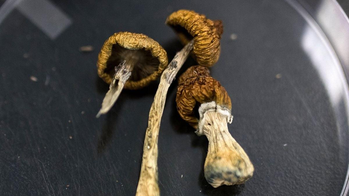
Now, a historically vilified psychedelic is emerging as a possible new avenue for controlling Alzheimer’s symptoms.
Neuroscientists around the world are starting to investigate if psilocybin – the psychoactive ingredient in magic mushrooms – can help protect the aging brain.
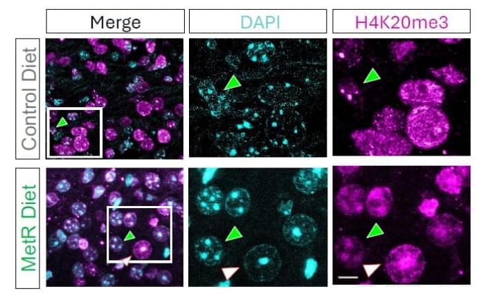
An unexpected lab observation has led a team of scientists to discover how diet can influence survival in animal models of glioma, one of the most aggressive and deadly forms of brain cancer. Researchers at Baylor College of Medicine, the Duncan Neurological Research Institute (Duncan NRI) at Texas Children’s Hospital and collaborating institutions report in the Proceedings of the National Academy of Sciences how limiting a single nutrient, the amino acid methionine, in the diet destabilized DNA organization and led to cancer cell death and increased animal survival. These findings open new possibilities for treating one of the most challenging forms of brain cancer.
“Cancer cells, including gliomas, often depend on methionine. Methionine is an essential amino acid, meaning that the body does not produce it on its own; it must be consumed in the diet. Glioma cells are unusually dependent on methionine to fuel rapid growth and control gene activity,” said corresponding author Dr. Benjamin Deneen, professor and Dr. Russell J. and Marian K. Blattner Chair in the Department of Neurosurgery and director of the Center for Cancer Neuroscience, all at Baylor.
“In the current study, we wanted to know, if tumors depend so much on methionine, what happens if we reduce the supply?” said first author Brittney Lozzi, a graduate student in the Deneen lab.


You may have heard the phrase “neurons that fire together wire together.” This short phrase summarizes the synaptic plasticity theory of learning described by Canadian psychologist Donald Hebb in his 1949 book The Organization of Behavior.
Hebb explained how the connections between neurons (brain cells) change as a result of repetitive firing. So when you repeat a movement like swinging a golf club over and over, the neural pathways involved in controlling that movement become stronger and faster. Not only do existing synapses (junctions between neurons) begin to fire more efficiently, but new synapses are formed and other neurons are recruited to get in on the action. As a result, your golf swing becomes more automatic, reliable, and forceful the more often you practice.
That is neuroplasticity: your brain’s ability to change and adapt based on input and use. The concept of neuroplasticity had been previously proposed by others, most notably American psychologists William James and Karl Lashley, and Polish neuroscientist Jerzy Konorski, but it was largely ignored by the scientific community until Hebb brought the concept to the forefront in his groundbreaking book.
This is a new ~1 hour talk by me on the concept of diverse intelligence, and morphogenesis as a model system with which to practice identifying and communicating with unconventional minds. This is a bit different than previous ones because I explicitly go over examples of how some of our various data on bioelectrics addresses each of several key properties of cellular collectives as cognitive agents navigating anatomical morphospace.
She had not spoken a full sentence in five years. Then she took a single 5 gram dose of psilocybin (which is a very large dose). She slept 19 hours. When she woke up, she spoke for hours about her life. She recognized family and held real conversations. She regained bladder control after five years, and walked on her own. She dressed herself. These gains continued for weeks.
Background:
Advanced Alzheimer’s disease (AD) is generally regarded as a stage of irreversible functional decline. Psilocybin is known to transiently alter large-scale brain network dynamics and to induce plasticity-related mechanisms in preclinical models, yet clinical data in advanced dementia remain lacking.
Case presentation:
We report the case of an octogenarian Japanese-American woman with a 10-year history of Alzheimer’s disease, including 5 years of marked hypofunction and predominantly monosyllabic speech. Baseline features included chronic urinary incontinence, executive dysfunction, dysphagia, dependent mobility, flat affect, and severe reduction in spontaneous communication. The patient received 5 g of orally administered psilocybin-containing mushrooms (Enigma strain). The acute phase was marked by autonomic activation, clinically suspected hyperthermia, profuse sweating, and a prolonged deep sleep-like state. Approximately 19 h post-administration, spontaneous autobiographical speech emerged.

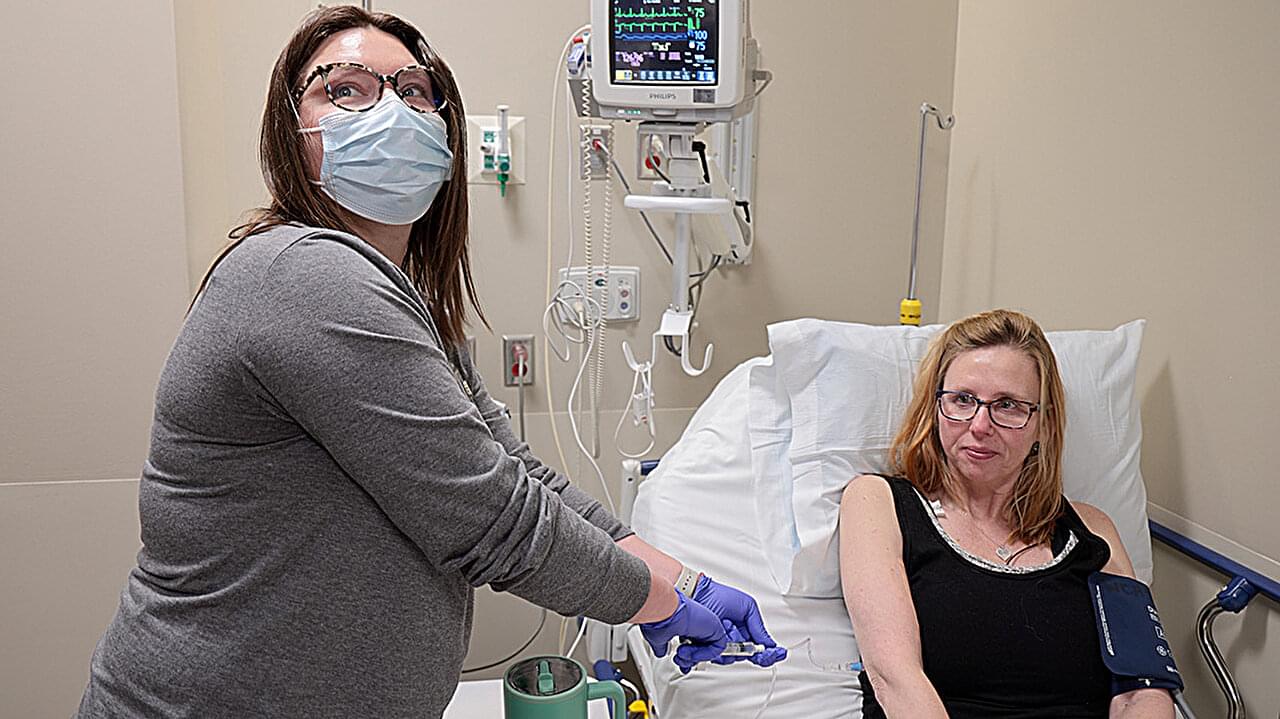
At age 49, Jan Janisch-Hanzlik’s multiple sclerosis was destroying her freedom to live the life she wanted. She gave up her active nursing job for a desk role. Frequent falls made her afraid to carry her grandchildren. She had to move to a bigger house to make room for the wheelchair she feared she might end up needing full-time.
Even the best available medication wasn’t improving Janisch-Hanzlik’s symptoms, and she worried they’d only get worse. So when she learned about a trial of CAR T cell therapy at the University of Nebraska Medical Center in Omaha, close to the city of Blair where she lives, she phoned the clinic every other month until they were ready to enroll her as the first patient.
Originally designed to target and wipe out cancer by reprogramming the patient’s immune cells, CAR T is now being offered to patients in hundreds of clinical trials for autoimmune conditions like multiple sclerosis, lupus, Graves’ disease, vasculitis and many others. The hope is that CAR T can duplicate the success it has demonstrated in a range of blood cancers by hunting down and eliminating cells that target the self in autoimmune diseases. This would essentially reset the body’s defenses to a state like the one that existed before the disease took hold.