A new study shows that faulty mitochondria may be a root cause of dementia symptoms. Stimulating these cellular “powerhouses” in mice restored memory, offering a potential new approach to treating neurodegenerative diseases.
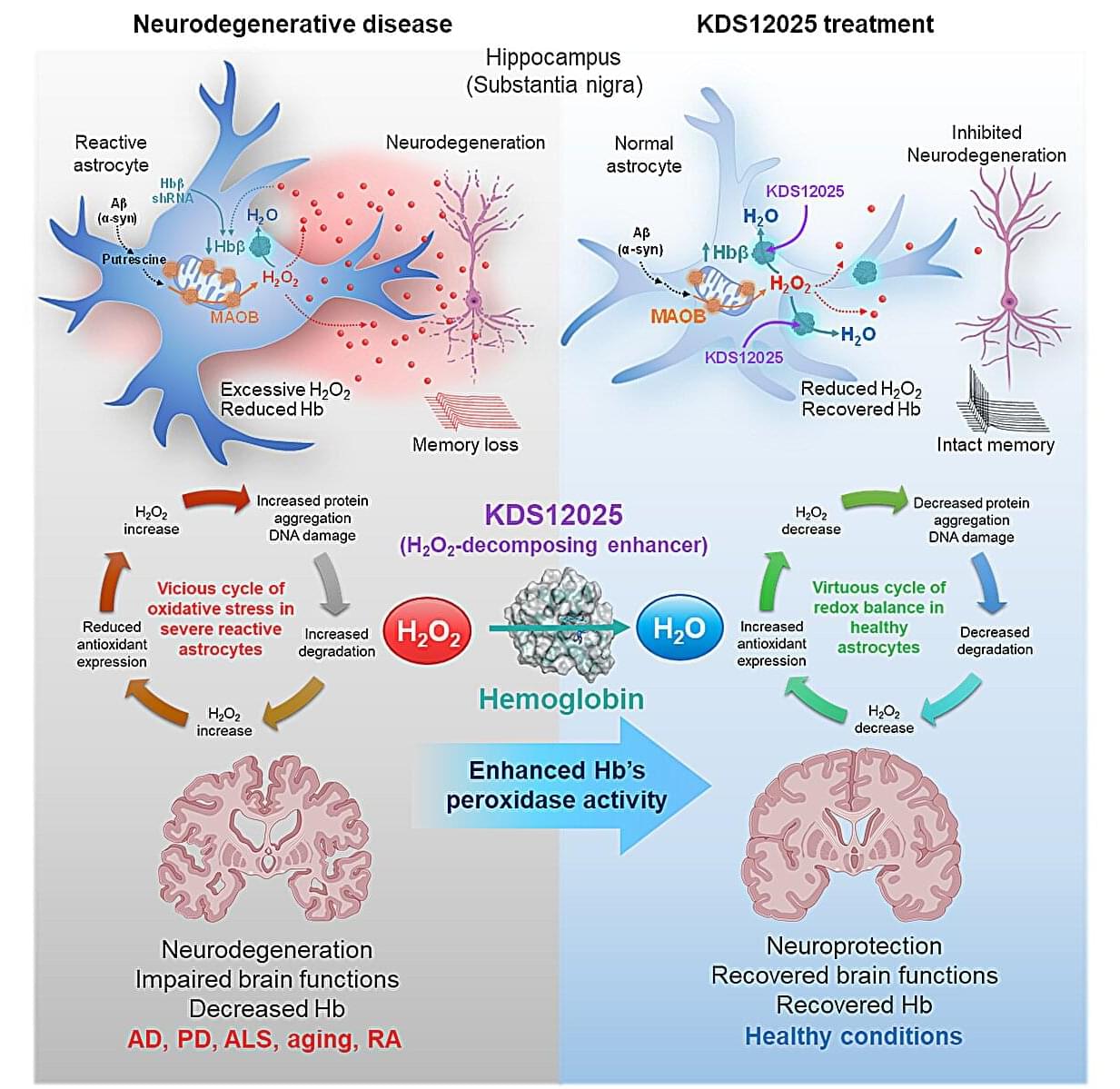
Hemoglobin, long celebrated for ferrying oxygen in red blood cells, has now been revealed to play an overlooked—and potentially game-changing—antioxidant role in the brain.
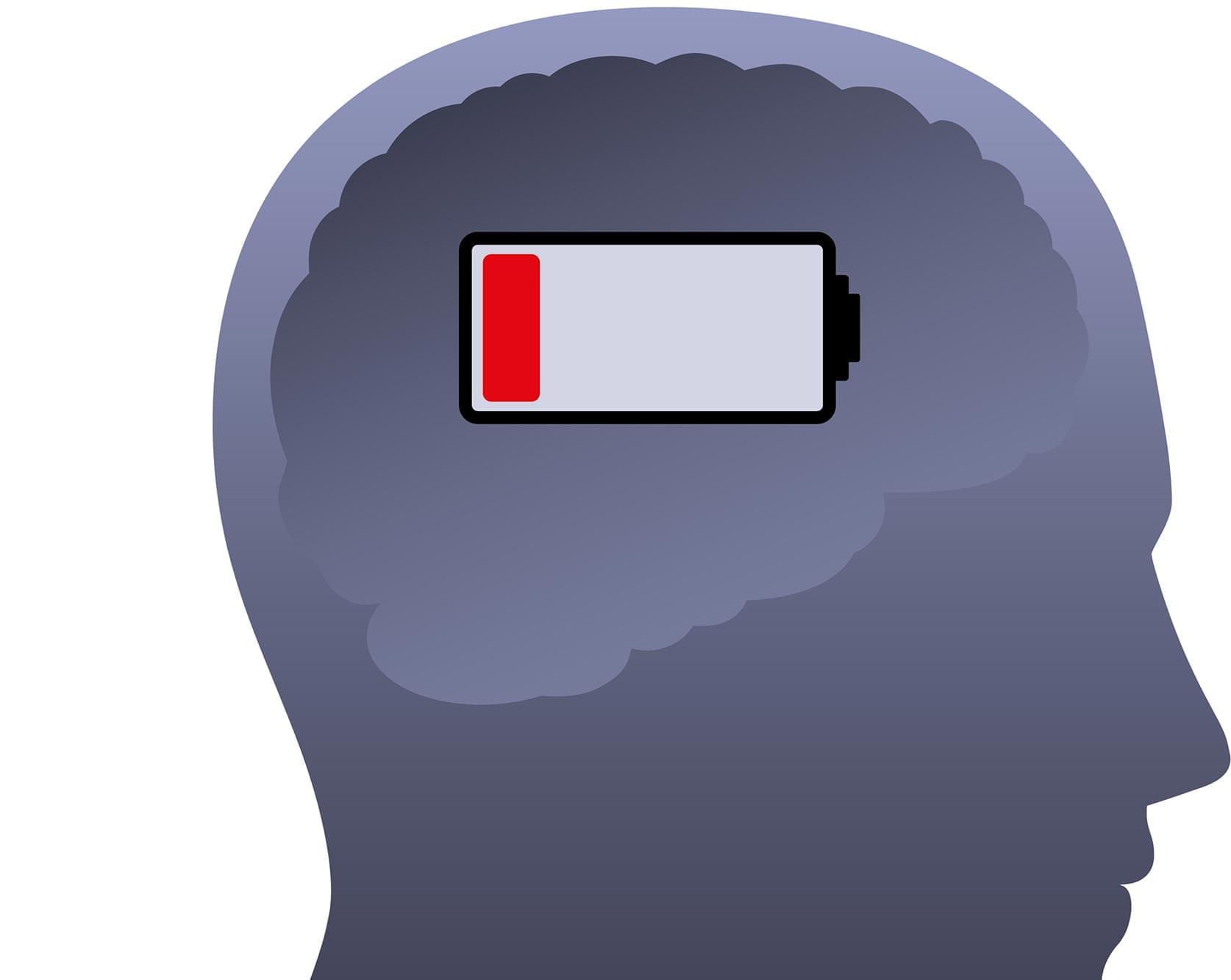
In neurodegenerative diseases such as amyotrophic lateral sclerosis (ALS), Parkinson’s, Alzheimer’s, and aging, brain cells endure relentless damage from the aberrant (or excessive) reactive oxygen species (ROS). For decades, scientists have tried to neutralize ROS with antioxidant drugs, but most failed: they couldn’t penetrate the brain effectively, were unstable, or indiscriminately damaged healthy cells.
This new study, led by Director C. Justin Lee of the Center for Cognition and Sociality within the Institute for Basic Science (IBS) in Daejeon, South Korea, set out to identify the brain’s own defenses against a particularly harmful form of ROS—hydrogen peroxide (H2O2). The study has been published in Signal Transduction and Targeted Therapy.

After living with psychiatric illnesses, including depression and PTSD, for many years and experiencing his first panic attacks when he was just a kindergartner, the patient in this study had been hospitalized numerous times. The authors write that he had endured “one protracted depressive episode without distinct periods of remission for 31 years.”
They describe his medical history as “remarkable” – he has tried at least 19 different medications and undergone electroconvulsive therapy (ECT) three times. While this treatment can be effective in some cases, in this patient it unfortunately left him with cognitive impairment.
Ultimately, the patient had experienced suicidal ideation and made attempts to take his own life. It’s thought that around a third of patients with major depressive disorder will progress to TRD, as in this case, and that is a strong risk factor for suicidality.

UNSW Sydney and Macquarie University psychology researchers have written an article warning that psychedelic therapies may switch on visual mental imagery in people with aphantasia and could raise the risk of intrusive thoughts, while calling for more detailed informed consent.
Known as a blind mind’s eye, people with aphantasia recall personal memories with fewer details and vividness. Visual mental imagery is absent. People with aphantasia cannot visualize objects, people, places, or memories, and they also recall personal memories with fewer details and vividness.
Recent reports, including one published case study and one pre-print along with anecdotal accounts, describe individuals with aphantasia gaining a new capacity to visualize after a single dose of ayahuasca or psilocybin, with positive self-reported outcomes during and after the experience, including within a year post-experience.