A new study reveals that the pace of a child’s brain maturation can predict whether they will tend to bottle up their emotions during their teenage years, offering new clues about the biological roots of adolescent mental health.

The report, titled “Pathways to Longevity”, introduces several important longevity concepts to the general reader and is another sign that the field is coming of age and entering the mainstream.
People do want to live longer
From time to time, Harvard Health Publishing issues Special Health Reports – consumer-facing, doctor-reviewed guides translating medical research for general readers. Previous reports included topics such as Alzheimer’s and heart disease. This new one, presented to the public earlier this week, is dedicated to healthy longevity. While this report, aimed mostly at curious laypeople and priced at $29, might not reveal a trove of new information to a longevity-savvy reader, it is an unmistakable sign that longevity science and the very idea of extending lifespan and healthspan are finally entering the mainstream.
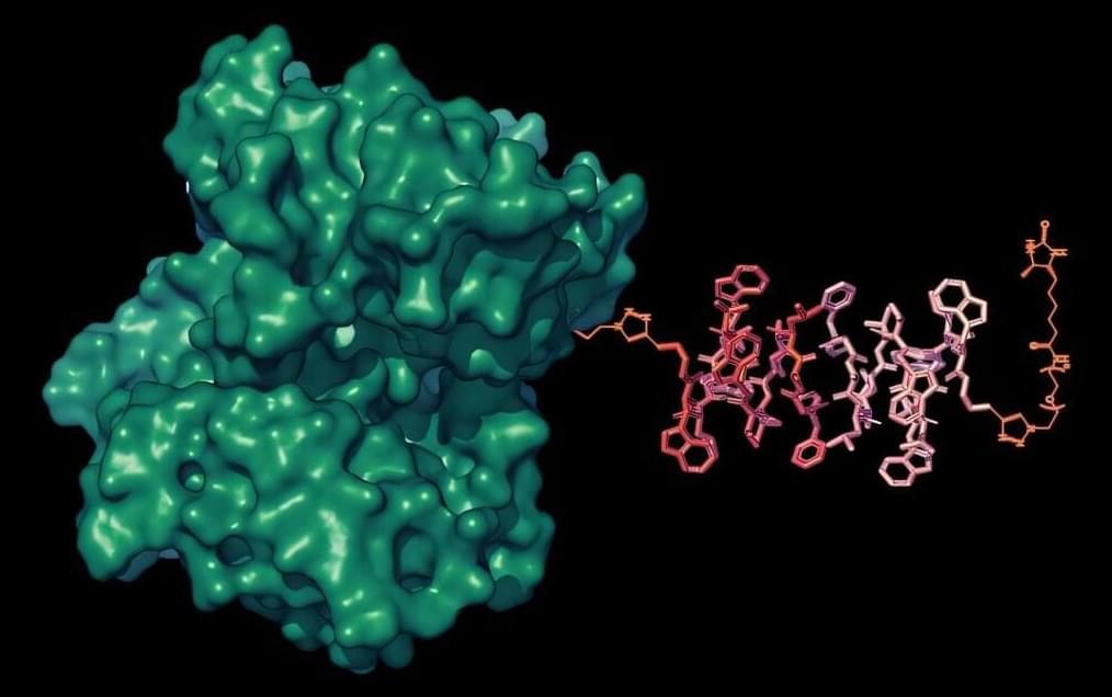
A newly identified protein called GPNMB may play a major role in helping Parkinson’s disease spread through the brain. Researchers discovered that immune cells release the protein in response to damaged neurons, creating a vicious cycle that speeds up brain cell degeneration. In early experiments, antibodies that blocked GPNMB stopped the toxic process from spreading between cells.
Investigadores enseñan a las células cerebrales a jugar a ‘Doom’
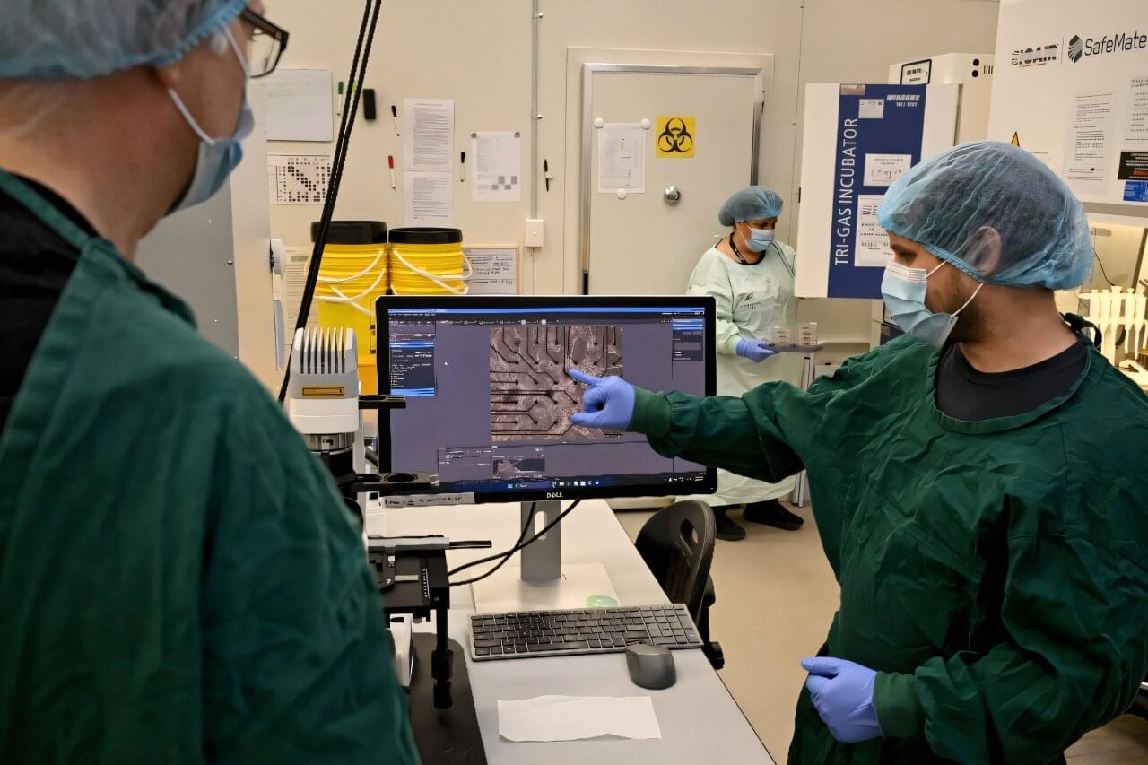
Australian researchers have trained lab-grown brain cells on a silicon computer chip to play the nineties shooter game “Doom” and say they are just scratching the surface of what the neurons could be capable of doing.
It’s the science-fiction work of biotech boffins at Cortical Labs, who researched and developed the technology that harnesses the workings of the brain’s networking system.
Each so-called “biological computer” contains around 200,000 living human brain cells, grown from stem cells that were harvested from blood donations.
In this exciting episode, we dive deep into the world of bio-inspired robotics with Prof. Auke Jan Ijspeert, a Swiss-Dutch roboticist and neuroscientist at the École Polytechnique Fédérale de Lausanne (EPFL). As the head of the Biorobotics Laboratory, Prof. Ijspeert shares how nature serves as the ultimate blueprint for designing the robots of the future. 🌿🤖
🔑 Key Highlights:
Bio-Inspired Robotics: Explore how Prof. Ijspeert and his team are mimicking nature to create innovative robots that move and behave like animals.
Neuroscience & Robotics: Learn how insights from neuroscience help reverse-engineer the sensorimotor coordination found in animals, applying it to robotic systems.
From Simulation to Reality: Discover the challenges of translating robotic simulations into real-world applications.
Exoskeletons & Assistive Technologies: Prof. Ijspeert discusses the development of exoskeletons for healthcare and military use, along with assistive furniture for people with limited mobility.
Humanoid Robots & Autonomous Systems: Get a sneak peek into the future of autonomous robotics, from central pattern generators to humanoid robots.
💡 Why You Should Watch:
Prof. Ijspeert is a trailblazer in the field of biorobotics, blending biology, neuroscience, and engineering to push the boundaries of what robots can achieve. Whether you’re a robotics enthusiast, a neuroscientist, or just curious about how nature inspires technology, this episode is packed with insights that could shape the future of robotics and artificial intelligence.
🔗 Connect with Prof. Auke Ijspeert:
https://www.epfl.ch/labs/biorob/peopl… / biorob_epfl
/ biorob_epfl Time Stamp 0:00 to 02:35 — Intro, Bio-Inspired Robots 02:35 to 04:13 — Neuroscience to back engineer bio-robots 04:13 to 06:22 — Mimicking nature & biorobots examples 06:22 to 07:55 — Simulation to real life translation challenges 07:55 to 09:10 — Central pattern generators & their role in robotic motion 09:10 to 10:47 — Learnings from creating bio-inspired robots 10:47 to 13:40 — EPFL Bio-Robotics laboratory 13:40 to 15:43 — Applications of Bio-robotics 15:43 to 20:05 — Exoskeleton 18:19 to 20:05 — Assertive furniture robotics 20:05 to 26:30 — exoskeleton in healthcare & military warfare 26:30 to 31:51 — Humanoid Robots 31:51 to 34:42 — Autonomous Robots 34:42 to 37:04 — Rhex Robots & Partnerships 37:04 to 40:04 — The future of robotics Watch our highest-viewed videos: 1-DR R VIJAYARAGHAVAN — PROF & PRINCIPAL INVESTIGATOR AT TIFR India’s 1st Quantum Computer–
• Quantum computer from India with Dr r vija… 2-TATA MOTORS-DRIVING THE FUTURE OF MOBILITY IN INDIA-SHAILESH CHANDRA-MD: TATA MOTORS–
• TATA MOTORS-DRIVING THE FUTURE OF MOBILIT… 3-MIT REPORT PREDICTS SOCIETAL COLLAPSE BY 2040 — GAYA HERRINGTON DIR SUSTAINABILITY: KPMG
• MIT Report predicts Total Societal Collaps… 4-WORLDS 1ST HUMAN HEAD TRANSPLANTATION-DR SERGIO CANAVERO —
• WORLDS 1ST HUMAN HEAD TRANSPLANTATION-DR… 5-DR HAROLD KATCHER — CTO NUGENICS RESEARCH Breakthrough in Age Reversal–
• BREAKTHROUGH IN AGE REVERSAL WITH YOUNGBLO… 6-How Neuroscience Will Change The Future Of Technology — Dr. James Giordano
• How Neuroscience Will Change The Future Of… 7-STARTUP FROM INDIA AIMING FOR LEVEL 5 AUTONOMY — SANJEEV SHARMA CEO SWAAYATT ROBOTS —
• SELF-DRIVING STARTUP FROM INDIA AIMING FOR… 8-MAN BEHIND GOOGLE QUANTUM SUPREMACY — JOHN MARTINIS —
• MAN BEHIND GOOGLE QUANTUM SUPREMACY — JOHN… 9-BANKING 4.0 — BRETT KING FUTURIST, BESTSELLING AUTHOR & FOUNDER MOVEN —
• BANKING 4.0 — BRETT KING FUTURIST, BESTSEL… 10-E-VTOL & HYPERLOOP-FUTURE OF INDIA” S MOBILITY-SATYANARAYANA CHAKRAVARTHY
• E-VTOL & HYPERLOOP-FUTURE OF INDIA“S MOBI… 11-HOW NEUROMORPHIC COMPUTING WILL ACCELERATE ARTIFICIAL INTELLIGENCE — PROF SHUBHAM SAHAY-IIT KANPUR–
• HOW NEUROMORPHIC COMPUTING WILL ACCELERATE… 12-How India Is Building a Quantum Computer — Dr. Anirban Bandyopadhyay
• How India Is Building a Quantum Computer -… Connect & Follow us at:
/ eddieavil
/ change-transform-india
/ changetransformindia
/ intothechange
/ changetransformindia Listen to the Audio Podcast at: https://anchor.fm/transform-impossible https://podcasts.apple.com/us/podcast… https://open.spotify.com/show/56IZXdz… https://www.breaker.audio/change-i-m–… https://www.google.com/podcasts?feed=… Don’t Forget to Subscribe
/ @toctwpodcast #robot #robotics #artificialintelligence #epfl.
In this video, we cover:
The Biological vs. The Synthetic: How the human brain’s hyper-dynamic, self-rewiring structure compares to the rigid \.
People who carry the APOE2 version of the apolipoprotein E gene are more likely to live to advanced age and are partly protected against Alzheimer’s disease, but scientists have struggled to explain why. A new study from the Buck Institute for Research on Aging, now published in Aging Cell, offers a mechanistic answer: APOE2 helps human neurons keep their DNA intact and resist becoming senescent, a damaged, dysfunctional state that accumulates with age and contributes to neurodegeneration.
The findings shift attention away from APOE’s well-known role in cholesterol transport and toward a previously underappreciated function of the gene: shaping how brain cells maintain the integrity of their genome as they age.
“We’ve known for years that APOE2 carriers tend to live longer and have a lower risk of Alzheimer’s, but the protective mechanism has been a black box,” says senior author Lisa M. Ellerby, Ph.D., professor at the Buck Institute. “Our work shows that APOE2 neurons are better at preventing and repairing DNA damage, and they resist the cellular aging program that drives so much of late-life decline. Our findings point to entirely new therapeutic directions.”
When doctors and scientists want to see inside a body, magnetic resonance imaging (MRI) is a powerful tool. MRI can noninvasively capture detailed images of the body’s muscles, organs, and bones. It can monitor blood flow to generate a map of brain activity. And with new sensors developed by bioengineers at MIT, MRI can track the kinds of molecules that make our brains and bodies work.
In the May 13 issue of the journal Nature Biomedical Engineering, a team led by Alan Jasanoff, the Eugene McDermott Professor in the Brain Sciences and Human Behavior at MIT, reports on their new sensors, which can brighten or dim MRI signals in response to specific molecular targets. The probes are designed to amplify the effect that each target molecule has on MRI signal, dramatically improving sensitivity over previous small-molecule sensors.
Jasanoff, who is also an associate investigator at the McGovern Institute for Brain Research, says the approach his team used should enable the development of MRI sensors that detect neurotransmitters and other important molecules in the brain.
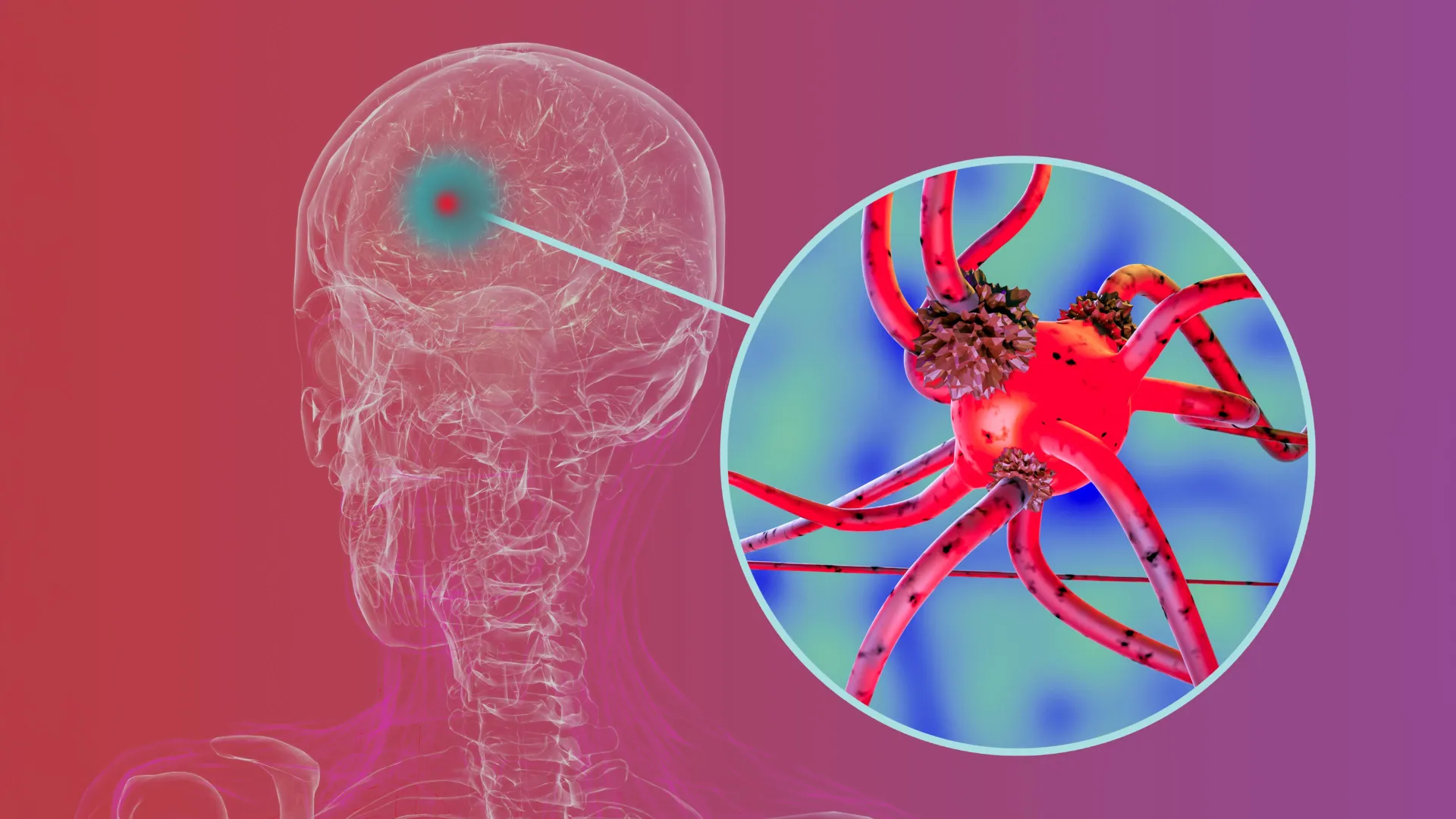
Researchers with the University of Cincinnati and Johns Hopkins Medicine developed a potential treatment for brain cancer that uses nanofibers embedded with a combination of drugs that work in concert to target tumors. The drugs proved more effective in combination than when administered alone and can provide both immediate and long-lasting doses to kill cancer cells.
“In our study, a three-drug combination showed strong synergistic effects across multiple glioblastoma models and significantly improved survival in animal studies,” said Daewoo Han, an assistant professor in UC’s College of Engineering and Applied Science and lead author of the paper published in ACS Biomaterials Science & Engineering.
Han and Distinguished Research Professor Andrew Steckl incorporated the drugs into electrospun fiber membranes, creating a nanofiber drug delivery system. Steckl’s NanoLab at the University of Cincinnati is a leading developer of this technology that uses an electric field to create a multilayered fiber mesh for drug delivery, among other uses. “This combination is pretty powerful,” Steckl said.
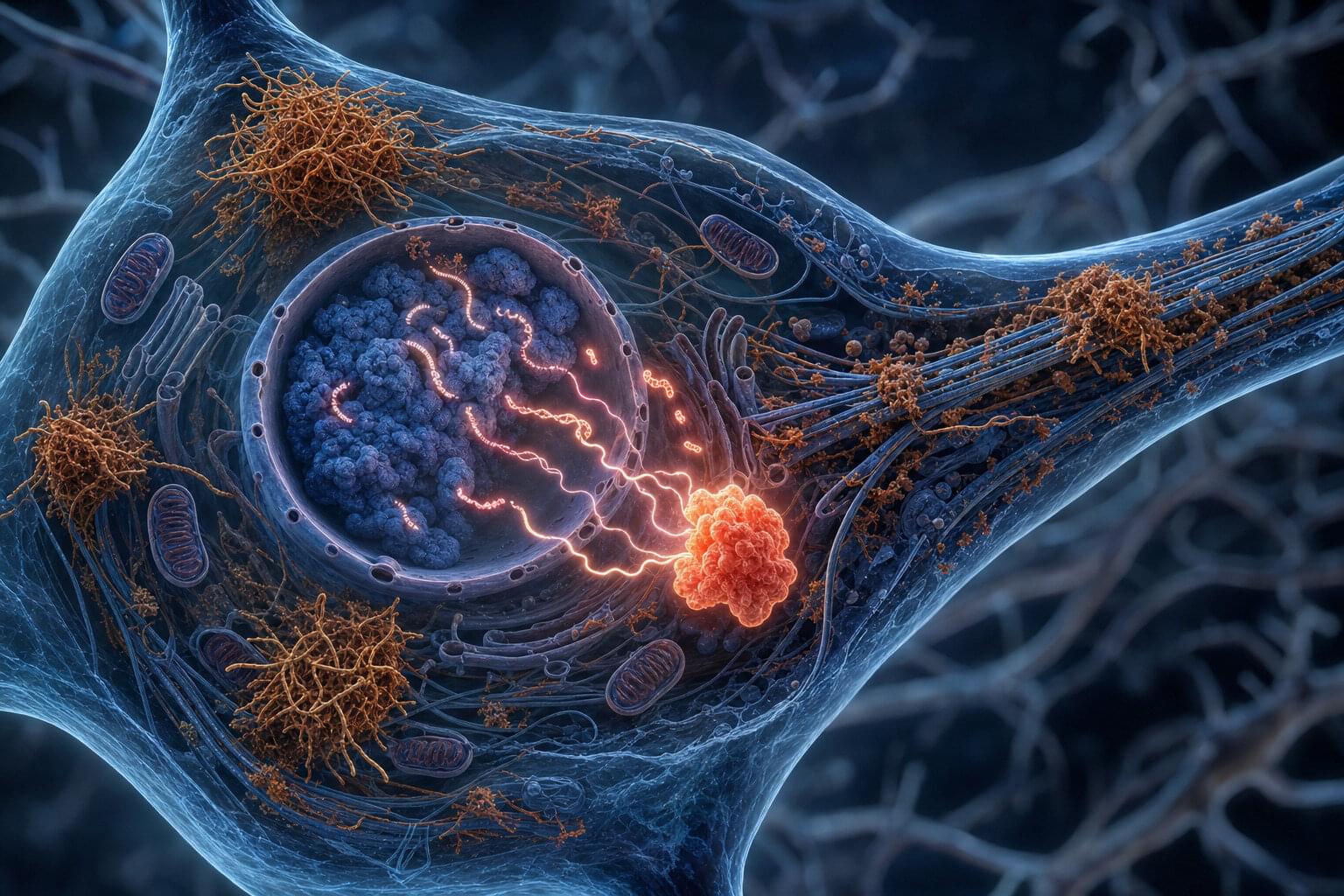
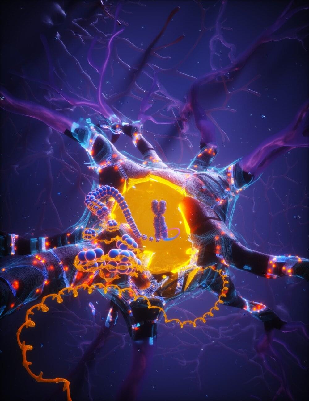
Alzheimer’s disease is a neurodegenerative disease characterized by a progressive decline in mental functions and memory loss. Along with frontotemporal dementia and some other neurodegenerative disorders, Alzheimer’s disease has been associated with an accumulation inside neurons of abnormal clumps of a protein called “tau.”
The tau protein is important for brain health, stabilizing structures called microtubules inside neurons. In Alzheimer’s disease and other tauopathies (i.e., diseases linked with the abnormal accumulation of tau), tau proteins aggregate into toxic and insoluble clumps that are harmful to brain cells, gradually leading to their death.
Researchers at Zhejiang University, Xiamen University and other institutes in China recently carried out a study aimed at better understanding the processes via which tau aggregation contributes to the death of neurons in patients with Alzheimer’s disease. Their findings, published in Nature Neuroscience, suggest that these tau clumps prompt the reactivation of transposable DNA elements in neurons, which can in turn lead to their death.