Study results suggest that cognitive health in later life is in part the product of lifetime exposure to cognitive enrichment.
Background and Objectives.
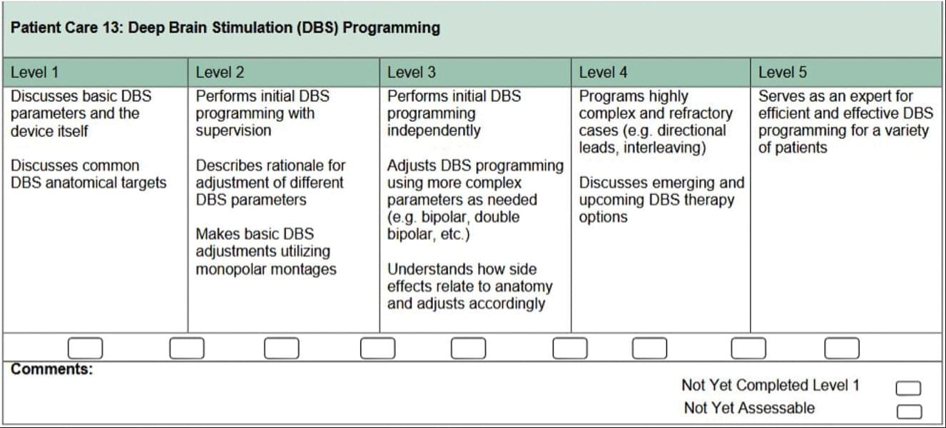
A Reflection on Movement Disorders —Fellowship Training in Deep Brain Stimulation: Past and Future.
Deep brain stimulation (DBS) has been an integral part of movement disorders care for decades. However, differences exist in techniques for surgical implantation of DBS and clinician experience with DBS systems, including use of new software, programming approaches, and postsurgical management of patients. DBS technologies have been rapidly advancing, and indications for DBS are increasing, including for psychiatric symptoms and epilepsy. The heterogeneity in the scope and utility of DBS is perhaps mirrored in education and training, despite efforts to develop competency measures for trainees. These advancements in DBS and the varying opportunities offered at each fellowship contribute to challenges for program directors to establish and implement consistent expectations. Similar challenges have been observed in other fields using neuromodulation.


UT Southwestern Medical Center researchers have identified two lipids that work together with a quintessential protein known as stimulator of interferon genes (STING) to launch an immune response in the human body. Their findings, detailed in two papers published concurrently in Nature, could lead to new ways to manipulate the immune system to fight infections, cancer, autoimmune disorders, and neurodegenerative diseases.
“These studies reveal additional levels of regulation of the cGAS-STING pathway, underscoring the importance of controlling the activity of this pathway so the body can mount an effective immune response against infections while avoiding autoimmune reactions to self-tissues. Dysregulation of this pathway has been shown to cause a variety of autoimmune and inflammatory diseases,” said Zhijian “James” Chen, Ph.D., Professor of Molecular Biology and Director of the Center for Inflammation Research at UT Southwestern.
Dr. Chen, one of the world’s leading researchers on innate immunity, is a co-author on one study and senior author on the other. His discovery of cGAS, an enzyme that produces a molecule called cGAMP to activate STING, has been recognized with numerous top honors including the 2026 Japan Prize in Life Sciences, the 2024 Albert Lasker Basic Medical Research Award, and the 2019 Breakthrough Prize in Life Sciences.
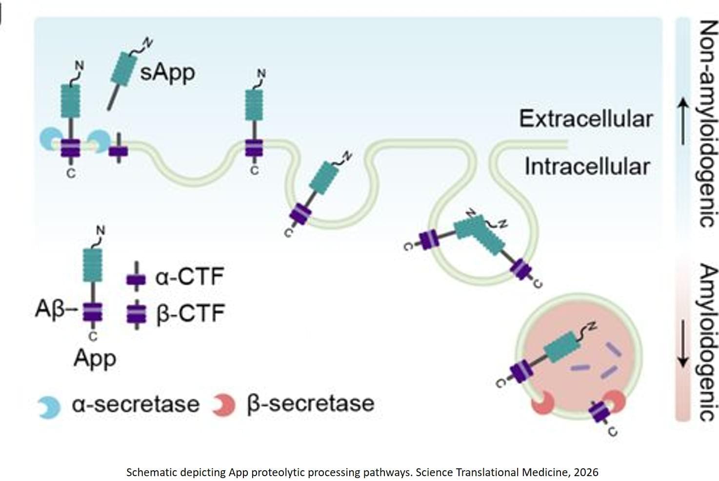
At the heart of the new discovery is amyloid precursor protein (APP), a protein that plays important roles in brain development and synaptic formation. Abnormal processing of APP can lead to the production of amyloid‑beta peptides, which play a central role in the development of Alzheimer’s disease. The scientists found that how APP is trafficked also controls whether a neuron forms amyloid-beta 42.
During the synaptic vesicle cycle — a fundamental process that underlies every thought, movement, memory or sensation — levetiracetam binds to a protein called SV2A. This interaction slows down a step in which neurons recycle synaptic vesicle components from the cell’s surface. By pausing this recycling process, the drug enables APP to remain on the cell’s surface longer, diverting it away from the pathway that produces toxic amyloid‑beta 42 proteins.
“In our 30s, 40s and 50s, our brains are generally able to steer proteins away from harmful pathways,” the author said. “As we age, that protective ability gradually weakens. This is not a statement of disease; this is just a part of aging. But in brains developing Alzheimer’s, too many neurons go astray, and that’s when you get amyloid-beta 42 production. And then it’s tau (or ‘tangles’), and then it’s dead cells, then dementia, then neuroinflammation — and then it’s too late.”
To effectively prevent Alzheimer’s symptoms, high-risk individuals would need to begin taking levetiracetam “very, very early,” the author said, possibly up to 20 years before the new FDA-approved Alzheimer’s disease test would even capture mildly elevated levels of amyloid-beta 42.
“You couldn’t take this when you already have dementia because the brain has already undergone a number of irreversible changes and a lot of cell death,” the author said.
Leveraging its status as an FDA-approved and widely used drug, the team mined existing human clinical data to investigate whether Alzheimer’s patients who took levetiracetam experienced slowed cognitive decline. They obtained clinical data from the National Alzheimer’s Coordinating Center and conducted a correlative analysis, finding that Alzheimer’s patients who took levetiracetam were associated with a significant delay from the diagnosis of cognitive decline to death compared to those taking lorazepam or no/other anti-epileptic drugs. ScienceMission sciencenewshighlights.
In this epsiode of the Cryosphere Chat we discuss:
● The themes of this year’s Biostasis Summit.
● Our thoughts on Tomorrw Bio’s big announcement about longevity experts.
● Greg Fahy’s paper on ultrastructure preservation in vitrified brains.
Links:
Buy tickets for the Biostasis days at Vitalist Bay: https://vitalistbay.com/ (use code CRYOSPHERE20 for 20% off)
Biostasis Summit needs based discount application: https://forms.gle/4pR3r4uvXprc4mH99
Biostasis Summit pitch application: https://forms.gle/FQsqx9thLvryKteq8
Join the Biostasis Summit mailing list: https://www.globalcryonicssummit.com/
Survey of cryonicists: https://cryospherepress.substack.com/p/the-cryonics-survey-of-2022-part.
Cryonics Subreddit: https://www.reddit.com/r/cryonics/
Cryosphere Discord: https://discord.gg/ndshSfQwqz.
Cryosphere Substack: https://cryospherepress.substack.com/
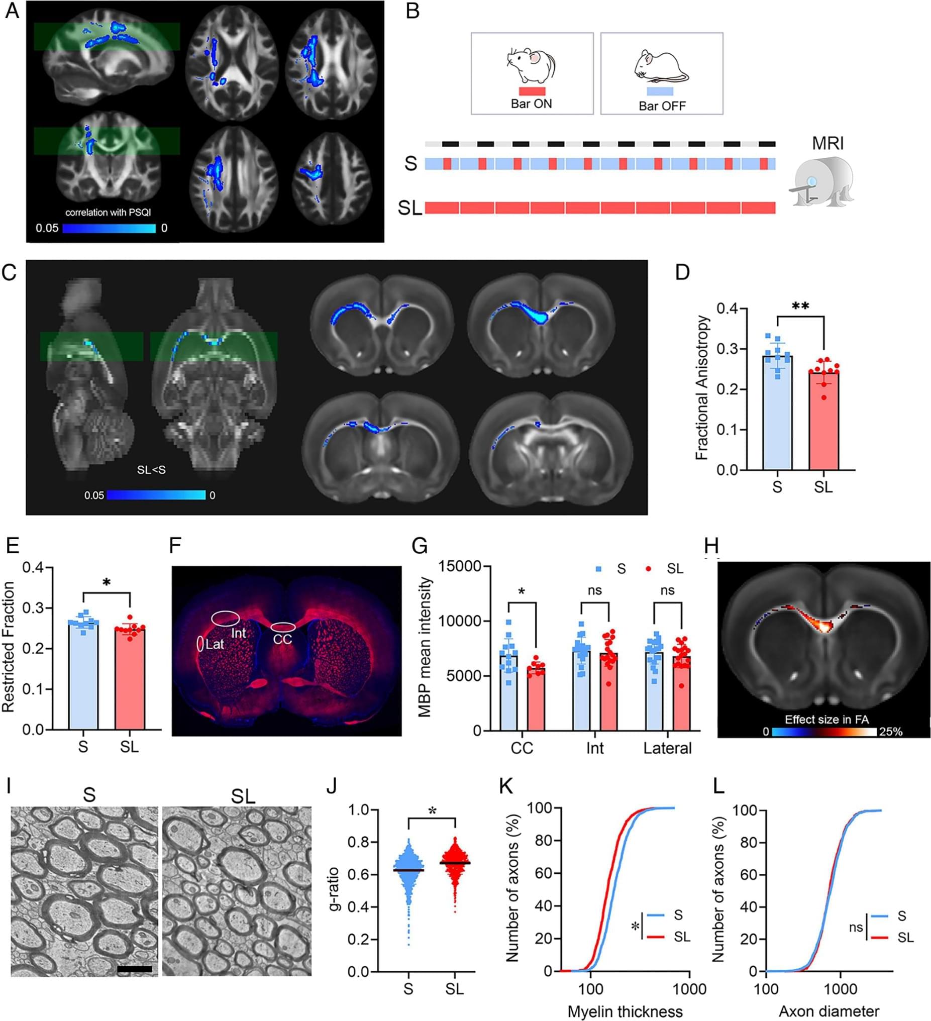
The increasing prevalence of sleep deprivation poses a public health challenge in modern society. Manifestations of reduced alertness, such as slowed reaction times and increased errors, are well-documented behavioral indicators of sleep loss (SL). Yet, the biological consequences of sleep deprivation and their role in behavioral impairment remain elusive. Our study reveals significant effects of sleep deprivation on myelin integrity. As a result, we identify increased conduction delays in nerve signal propagation, hindered interhemispheric synchronization, and impaired cognitive and motor performance associated with SL. By profiling oligodendrocyte transcriptome and lipidome, we observe SL-induced endoplasmic reticulum stress and lipid metabolism dysregulation, particularly affecting cholesterol homeostasis.

An Oregon State University scientist and a team of undergraduate students have uncovered real-time insights into a chemical process linked with Alzheimer’s disease, paving the way toward better drug designs. The researchers used a molecule measuring technique to observe in a laboratory setting how certain metals can promote the protein clumping that leads to the blocked neural pathways associated with Alzheimer’s. Led by Marilyn Rampersad Mackiewicz, associate professor of chemistry in the OSU College of Science, the research team also watched molecules known as chelators disrupt or reverse the clumping. The findings are published in ACS Omega.
Alzheimer’s disease is the most common form of dementia, a chronic condition of impaired cognitive function that affects large numbers of older adults and their loved ones. According to the Centers for Disease Control and Prevention, Alzheimer’s is the sixth-leading cause of death for people age 65 and older.
In Alzheimer’s patients, aggregations of amyloid-beta proteins interrupt brain cells’ ability to communicate with each other. The brain needs certain metals to work properly, but problems arise when the metals are present in unbalanced quantities.
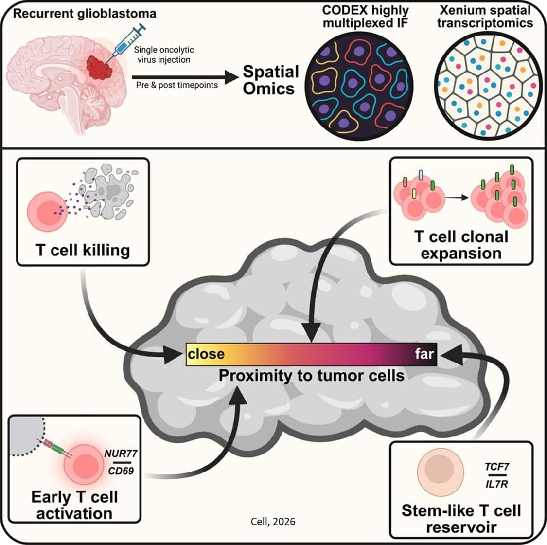
A team led by investigators has shown that a single injection of an oncolytic virus—a genetically modified virus that selectively infects and destroys cancer cells—can recruit immune cells to penetrate and persist deep within brain tumors. The research, which is published in Cell, provides details on how this therapy prolonged survival in patients with glioblastoma, the most common and malignant primary brain tumor, in a recent clinical trial.
The oncolytic virus used in the team’s trial is made from a herpes simplex virus genetically altered so it can only make copies of itself in glioblastoma cells and not normal healthy cells. The virus spreads to a glioblastoma cell, kills it, and then makes a copy of itself that spreads again to another glioblastoma cell. Infection of cells with the virus also triggers an immune response. In the phase 1 trial of 41 patients with recurrent glioblastoma, the oncolytic virus treatment extended survival compared to historically reported survival, especially among those with pre-existing viral antibodies.
In their Cell study, the investigators examined the extent of this immune response in clinical trial participants. Their analysis revealed that the treatment induced long-term infiltration of immune T cells into patients’ tumors. Closer proximity of cytotoxic T cells with dying brain tumor cells was associated with longer patient survival after treatment. The therapy also expanded pre-existing T cells in the brain. ScienceMission sciencenewshighlights.