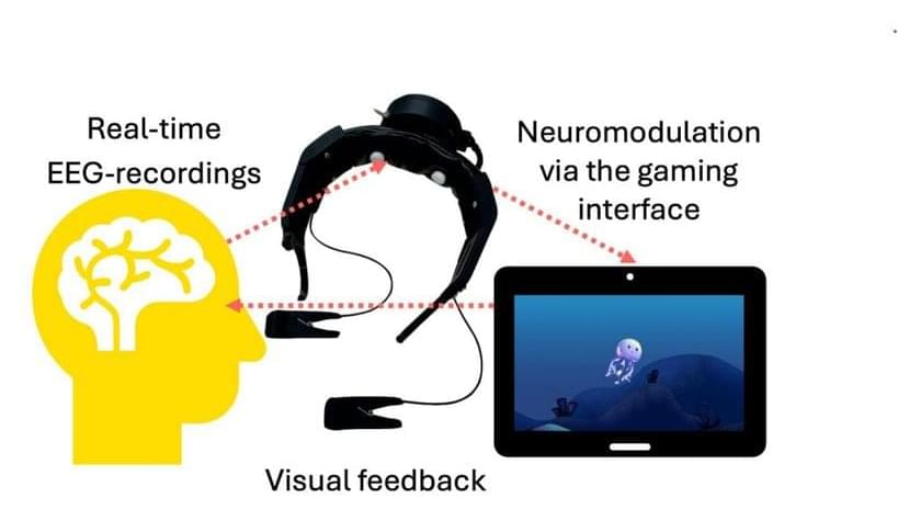
Toggle light / dark theme

We Can Now Simulate a Human Brain, Scientists Show

Go to https://ground.news/sabine to get 40% off the Vantage plan and see through sensationalized reporting. Stay fully informed on events around the world with Ground News.

Over the years, computer scientists have used cutting-edge processors to simulate the brains of increasingly more complex animals. They’ve already simulated worm and fruit fly brains, and are now working on mice. But according to a new paper, they’ve made a breakthrough that might allow them to simulate human brains, which contain 80 billion neurons compared to a fruit fly’s 140,000. Let’s take a look.

Paper: https://arxiv.org/abs/2512.

👕T-shirts, mugs, posters and more: ➜ https://sabines-store.dashery.com/
💌 Support me on Donorbox ➜ https://donorbox.org/swtg.
👉 Transcript with links to references on Patreon ➜ / sabine.
📝 Transcripts and written news on Substack ➜ https://sciencewtg.substack.com/
📩 Free weekly science newsletter ➜ https://sabinehossenfelder.com/newsle… Audio only podcast ➜ https://open.spotify.com/show/0MkNfXl… 🔗 Join this channel to get access to perks ➜ / @sabinehossenfelder 📚 Buy my book ➜ https://amzn.to/3HSAWJW #science #sciencenews #neuroscience #tech.
👂 Audio only podcast ➜ https://open.spotify.com/show/0MkNfXl
🔗 Join this channel to get access to perks ➜
/ @sabinehossenfelder.
📚 Buy my book ➜ https://amzn.to/3HSAWJW

#science #sciencenews #neuroscience #tech

Mitochondria-Associated MicroRNAs: Emerging Roles in the Pathogenesis of Parkinson’s Disease

💡 by Catanesi, M., et al. (2026). Biomedicines, 14, 313. 📖Read the full text: https://brnw.ch/21x1AQl.

Neurodegenerative diseases continue to challenge modern medicine, with mitochondrial dysfunction emerging as one of their most critical hallmarks. Among the most intriguing recent discoveries is the role of mitochondrial‑localized microRNAs (mito‑miRNAs), small regulatory molecules capable of influencing mitochondrial gene expression and cellular metabolism. As research uncovers their presence and function within the mitochondrial environment, these molecules are gaining attention for their potential involvement in the onset and progression of disorders such as Parkinson’s disease. Understanding how mito‑miRNAs contribute to neuronal vulnerability may open new avenues for diagnostics and therapeutic strategies in neurodegeneration.


Neurodegenerative diseases (NDs) are the most prevalent age-associated disorders, characterized by progressive neuronal loss and cognitive decline. Mitochondrial dysfunction is strictly associated with NDs and represent one of the hallmarks of these disorders, with neurological syndromes frequently representing the primary clinical manifestations of mitochondrial abnormalities. As central regulators of cellular bioenergetics, mitochondria play a pivotal role in both the physiological maintenance and pathogenesis of disease by different regulatory approaches. One of these, microRNAs (miRNAs), a class of small non-coding RNAs, are well-established regulators of gene expression across different biological pathways.

A missing link in how the brain regulates appetite

Bile acids at the center of hepato-ocular crosstalk.

Hepatic dysfunction with ocular pathology has been linked to dysregulated bile acid metabolism.

Bile acid imbalance has been shown to drive ocular injury along the gut-liver-eye axis through direct cytotoxicity, disruption of retinal and lens homeostasis mediated by FXR and TGR5 signaling, and immune activation and these mechanisms are implicated across a spectrum of conditions, ranging from inborn metabolic disorders to acquired cholestatic diseases.

The researchers in this review discuss translational potential of targeting bile acid homeostasis and summarize emerging therapeutic strategies, including bile acid-based interventions, targeted drug delivery, and microbiome modulation, that aim to restore systemic bile acid balance.

Thus, bile acid homeostasis act as a unifying therapeutic framework for hepato-ocular comorbidities. sciencenewshighlights ScienceMission https://sciencemission.com/Hepato-ocular-crosstalk


Health sciences.

This nasal spray rewinds the aging brain, restoring memory and reversing inflammation in preclinical models

Picture this: your brain is a high-performance engine. Over decades, it doesn’t just wear down, it also starts to run hot. Tiny “fires” of inflammation smolder deep within the brain’s memory center, creating a persistent brain fog that makes it harder to think, form new memories or even adapt to new environments, all the while increasing the risk to disorders like Alzheimer’s disease.

Scientists call this slow burn “neuroinflammaging,” and for decades it was thought to be the inevitable price of growing older. Until now.

A landmark study by researchers at Texas A&M University Naresh K. Vashisht College of Medicine suggests the inflammatory tide responsible for brain aging and brain fog might actually be reversible. And the solution doesn’t involve brain surgery, but a simple nasal spray.

Perovskite Nanocrystals, Quantum Dots, and Two-Dimensional Structures: Synthesis, Optoelectronics, Quantum Technologies, and Biomedical Imaging

💬Editorial: Decade-long follow-up highlights that patients with surgically treated ChronicSubduralHematoma have persistent excess mortality and long-term cognitive and functional impairment, even when overall quality of life appears preserved.


In the setting of aging populations and rising antithrombotic use, chronic subdural hematomas (cSDH) are increasingly common in high-income countries. While discussed in the medical literature for centuries,1 clinical research on cSDH in the modern era initially focused on surgical approaches and the risk of short-term mortality and recurrence.2-4 In this setting, cSDH was perceived to be a relatively benign disease; however, recent work has challenged this reputation. Patients with cSDH have persistently elevated long-term mortality when compared with controls,5 and those who survive are often left with functional and cognitive impairment.6 Unfortunately, most prior studies of long-term outcomes were small or had limited data on premorbid health, and none had data on functional status or quality of life.

In this issue of JAMA Neurol ogy, Petutschnigg et al7 expand on this work by examining mortality, function, and quality of life 10 years after surgical management of cSDH. To achieve this, they used patients who had previously been enrolled in a cSDH clinical trial from 2012 through 2016, which was conducted at a single center in Switzerland and examined the use of routine follow-up computed tomography scans in surgically managed patients with cSDH. The study authors obtained all-cause mortality for all 359 participants through 2023 using a nationwide data source. Each patient was then matched by age, sex, and birth month to controls from the Swiss population. Then, they obtained health-related quality of life by administering a validated survey to consenting participants, getting a response in 147 of 202 survivors at a mean of 10.55 years from the cSDH. Results from these participants were compared with normative values for a European population, using standardized age and sex strata.

The authors7 found a significantly higher mortality rate among patients with cSDH when compared with controls, with the absolute risk difference widening from 6% at 1 year to 18% at 10 years. Among those who survived, both men and women showed significant impairment in cognitive and role functioning (ie, how much their daily work/hobbies are impaired) when compared with normative controls. In addition, men (but not women) showed significant additional impairment in physical functioning and social functioning when compared with normative controls. Importantly, perceived quality of life was not reduced in either men or women. Discordance between functional impairments and perceived quality of life has been observed in other types of brain injury, a phenomenon termed the disability paradox,8 and should similarly caution against therapeutic nihilism when it comes to patients with cSDH.

Scientists Grow Electronics Inside the Brains of Living Mice

Yet most implants require extensive surgery and risk damaging the brain’s delicate tissue. The new technology would avoid these downsides by building electrodes directly at the target.

“Our work points to a future where doctors could ‘grow’ soft, wire-free electronic interfaces inside the brain using the patient’s own blood, then gently dial brain activity up or down from outside the head using harmless near-infrared light,” study author Krishna Jayant said in a press release.

The brain produces every one of our sensations, movements, emotions, and decisions. Scientists have long sought to decode and manipulate its activity with a range of hardware.

Nociceptive neurons protect cancer cells against oxidative stress

How cancer cells exploit the tumor microenvironment to alleviate oxidative stress remains largely unclear. Zhang et al. find that nociceptive neurons, via secretion of EREG, protect HNSCC against oxidative stress-induced cell death. Targeting nociceptive neurons improves the therapeutic efficacy of chemotherapies, including cisplatin.

/* */