Toggle light / dark theme

The quantum revolution: Brain waves

Presented by Madhumita Murgia and John Thornhill, produced by Josh Gabert-Doyon and Edwin Lane. Executive producer is Manuela Saragosa. Sound design by Breen Turner and Samantha Giovinco. Original music by Metaphor Music. The FT’s head of audio is Cheryl Brumley. Special thanks to The Hospital for Sick Children.

We’re keen to hear more from our listeners about this show and want to know what you’d like to hear more of, so we’re running a survey which you can find at ft.com/techtonicsurvey. It takes about 10 minutes to complete and you will be in with a chance to win a pair of Bose QuietComfort earbuds.

Read a transcript of this episode on FT.com.

Does consciousness arise from the brain? | Donald Hoffman challenges Hannah Critchlow

Donald Hoffman and Hannah Critchlow debate the origins of consciousness.

This excerpt was taken from “The key to consciousness,” featuring Sam Coleman, Donald Hoffman, and Hannah Critchlow. Joanna Kavenna hosts.

Watch the full debate at https://iai.tv/video/the-key-to-consciousness?utm_source=You…escription.

#MysteriousConsciousness #ConsciousnessUnlocked #IsConsciousnessMaterial.

Donald Hoffman is an American cognitive psychologist working at the University of California, Irvine. He is making waves with a new theory suggesting that, instead of presenting reality as it “really is”, our perception is like a desktop interface enabling us to use reality effectively.

Named as one of the ten leading “communicator scientists” in the UK by the Science Council, Hannah Critchlow is an internationally-acclaimed neuroscientist with a background in neuropsychiatry.

PiEEG Offers Affordable Brain-Computer Interface

One day in the future, we may interact with our electronic devices not with physical input or even voice commands, but simply by thinking about what we want to do. Such brain–computer interfaces (BCIs), combined with machine learning, could allow us to turn our ideas into reality faster and with less effort than ever before — imagine being able to produce a PCB design simply by thinking about how the completed circuit would work. Of course as an assistive technology, BCIs would be nothing less than life-changing for many.

Today BCIs are in their infancy, but that doesn’t mean there isn’t room for hackers and makers to experiment with the concept. [Ildar Rakhmatulin] has been working on low-cost open source BCIs for years, and with the recent release of his PiEEG on Crowd Supply, thinks he’s finally found an affordable solution that will let individuals experiment with this cutting edge technology.

Implemented as a shield that can be connected to a Raspberry Pi 3 or 4, the PiEEG features 8 channels for connecting wet or dry electrodes that can measure biosignals such as those used in electroencephalography (EEG), electromyography (EMG), and electrocardiography (ECG). With the electrodes connected, reading these biosignals is as easy as running a Python script. While primarily designed for neuroscience experimentation, [Ildar] says the device is also useful for learning more about signal processing, filters, and machine learning.

The Synaptic Extracellular Matrix: Long-Lived, Stable, and Still Remarkably Dynamic

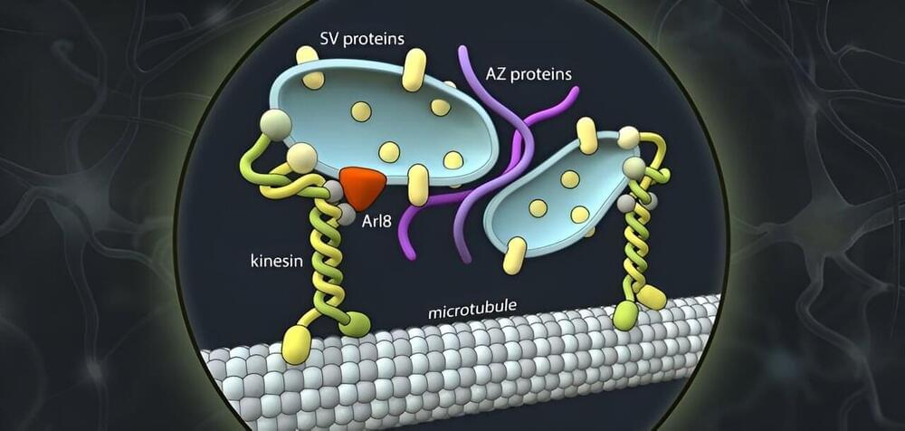
In the adult brain, synapses are tightly enwrapped by lattices of the extracellular matrix that consist of extremely long-lived molecules. These lattices are deemed to stabilize synapses, restrict the reorganization of their transmission machinery, and prevent them from undergoing structural or morphological changes. At the same time, they are expected to retain some degree of flexibility to permit occasional events of synaptic plasticity. The recent understanding that structural changes to synapses are significantly more frequent than previously assumed (occurring even on a timescale of minutes) has called for a mechanism that allows continual and energy-efficient remodeling of the extracellular matrix (ECM) at synapses. Here, we review recent evidence for such a process based on the constitutive recycling of synaptic ECM molecules. We discuss the key characteristics of this mechanism, focusing on its roles in mediating synaptic transmission and plasticity, and speculate on additional potential functions in neuronal signaling.

An increasing number of studies are showing that synaptic function is strongly influenced by their local environment, including the molecules or cellular components in their vicinity. As a result, the classical synaptic framework (consisting of the pre-and postsynaptic compartments only) has gradually been extended to include the neighboring astrocytic processes (the “tripartite synapse”; Araque et al., 1999) and, ultimately, also the surrounding extracellular matrix (ECM; the “tetrapartite synapse”; Dityatev et al., 2006). Nowadays, the synaptic ECM is recognized to play an essential role in physiological synaptic transmission as well as in plasticity, and its dysregulation has been linked to synaptopathies in a wide variety of brain disorders (Bonneh-Barkay and Wiley, 2009; Pantazopoulos and Berretta, 2016; Ferrer-Ferrer and Dityatev, 2018).

Alzheimer’s May Not Actually Be a Brain Disease, Expert Says

The pursuit of a cure for Alzheimer’s disease is becoming an increasingly competitive and contentious quest with recent years witnessing several important controversies.

In July 2022, Science magazine reported that a key 2006 research paper, published in the prestigious journal Nature, which identified a subtype of brain protein called beta-amyloid as the cause of Alzheimer’s, may have been based on fabricated data.

One year earlier, in June 2021, the US Food and Drug Administration had approved aducanumab, an antibody-targeting beta-amyloid, as a treatment for Alzheimer’s, even though the data supporting its use were incomplete and contradictory.

Fluid flow in the brain can be manipulated

Researchers at Boston University, U.S. report that the flow of cerebrospinal fluid in the brain is linked to waking brain activity. Led by Stephanie Williams, and publishing in the open access journal PLOS Biology on March 30, the study demonstrates that manipulating blood flow in the brain with visual stimulation induces complementary fluid flow. The findings could impact treatment for conditions like Alzheimer’s disease, which have been associated with declines in cerebrospinal fluid flow.

Just as our kidneys help remove from our bodies, cerebrospinal fluid helps remove toxins from the brain, particularly while we sleep. Reduced flow of cerebrospinal fluid is known to be related to declines in brain health, such as occur in Alzheimer’s disease. Based on evidence from , the researchers hypothesized that while awake could also affect the flow of cerebrospinal fluid. They tested this hypothesis by simultaneously recording human brain activity via fMRI and the speed of cerebrospinal fluid flow while people were shown a checkered pattern that turned on and off.

Researchers first confirmed that the checkered pattern induced brain activity; blood oxygenation recorded by fMRI increased when the pattern was visible and decreased when it was turned off. Next, they found that the flow of cerebrospinal fluid negatively mirrored the blood signal, increasing when the checkered pattern was off. Further tests showed that changing how long the pattern was visible affected blood and fluid in a predictable way, and that the blood-cerebrospinal fluid link could not be accounted for by only breathing or heart rate rhythms.

How Your Brain Organizes Information

To try everything Brilliant has to offer—free—for a full 30 days, visit http://brilliant.org/ArtemKirsanov/
The first 200 of you will get 20% off Brilliant’s annual premium subscription.

My name is Artem, I’m a computational neuroscience student and researcher. In this video we talk about cognitive maps – internal models of outside world that the brain to generate flexible behavior that is generalized across contexts.

Patreon: https://www.patreon.com/artemkirsanov.
Twitter: https://twitter.com/ArtemKRSV

OUTLINE:
00:00 — Introduction.
02:08 — Edward Tolman.
03:48 — Zoo of neurons in hippocampal formation.
06:40 — Non spatial mapping.
08:21 — Graph formalism.
12:21 — Latent spaces.
17:22 — Factorized representations.
21:51 — Summary.
24:47 — Brilliant.
26:19 — Outro.

REFERENCES (in no particular order):
1. Behrens, T. E. J. et al. What Is a Cognitive Map? Organizing Knowledge for Flexible Behavior. Neuron 100490–509 (2018).
2. Constantinescu, A. O., O’Reilly, J. X. & Behrens, T. E. J. Organizing conceptual knowledge in humans with a gridlike code. Science 352, 1464–1468 (2016).
3. Aronov, D., Nevers, R. & Tank, D. W. Mapping of a non-spatial dimension by the hippocampal–entorhinal circuit. Nature 543719–722 (2017).
4. Whittington, J. C. R., McCaffary, D., Bakermans, J. J. W. & Behrens, T. E. J. How to build a cognitive map. Nat Neurosci 25, 1257–1272 (2022).
5. Whittington, J., Muller, T., Mark, S., Barry, C. & Behrens, T. Generalisation of structural knowledge in the hippocampal-entorhinal system.

CREDITS:

/* */