How does the developing brain process surprising sounds and what changes as we grow up?
For children, the world is full of surprises. Adults, on the other hand, are much more difficult to surprise. And there are complex processes behind this apparently straightforward state of affairs. Researchers at the University of Basel have been using mice to decode how reactions to the unexpected develop in the growing brain.
Babies love playing peekaboo, continuing to react even on the tenth sudden appearance of their partner in the game. Recognizing the unexpected is an important cognitive ability. After all, new can also mean dangerous.
The exact way in which surprises are processed in the brain changes as we grow, however: unusual stimuli are much more quickly categorized as “important” or “uninteresting,” and are significantly less surprising the second and third time they appear. This increased efficiency makes perfect sense: new stimuli may gain our attention, but do not cause an unnecessarily strong reaction that costs us energy. While this may appear trivial at first, so far there has been very little research into this fact in the context of brain development.
In our new paper, we’ve investigated how quantum particles could move in a complex structure like the brain, but in a lab setting. If our findings can one day be compared with activity measured in the brain, we may come one step closer to validating or dismissing Penrose and Hameroff’s controversial theory.
Brains and Fractals
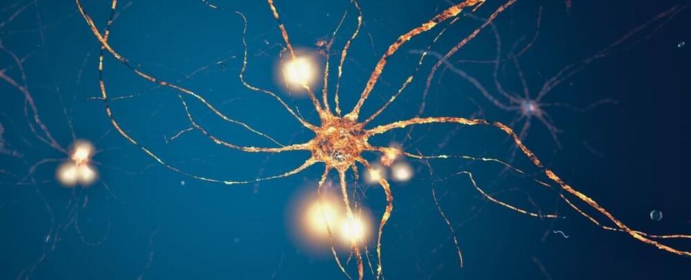
Our brains are composed of cells called neurons, and their combined activity is believed to generate consciousness. Each neuron contains microtubules, which transport substances to different parts of the cell. The Penrose-Hameroff theory of quantum consciousness argues that microtubules are structured in a fractal pattern which would enable quantum processes to occur.
Our memories are rich in detail: we can vividly recall the color of our home, the layout of our kitchen, or the front of our favorite café. How the brain encodes this information has long puzzled neuroscientists.
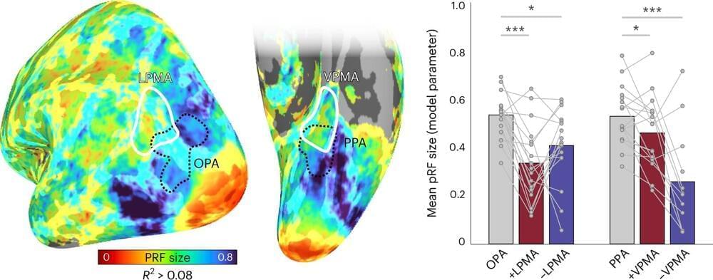
In a new Dartmouth-led study, researchers identified a neural coding mechanism that allows the transfer of information back and forth between perceptual regions to memory areas of the brain. The results are published in Nature Neuroscience.
Prior to this work, the classic understanding of brain organization was that perceptual regions of the brain represent the world “as it is,” with the brain’s visual cortex representing the external world based on how light falls on the retina, “retinotopically.” In contrast, it was thought that the brain’s memory areas represent information in an abstract format, stripped of details about its physical nature. However, according to the co-authors, this explanation fails to take into account that as information is encoded or recalled, these regions may in fact, share a common code in the brain.
Animals exhibit a diverse behavioral repertoire when exploring new environments and can learn which actions or action sequences produce positive outcomes. Dopamine release upon encountering reward is critical for reinforcing reward-producing actions1 – 3. However, it has been challenging to understand how credit is assigned to the exact action that produced dopamine release during continuous behavior. We investigated this problem with a novel self-stimulation paradigm in which specific spontaneous movements triggered optogenetic stimulation of dopaminergic neurons. Dopamine self-stimulation rapidly and dynamically changes the structure of the entire behavioral repertoire. Initial stimulations reinforced not only the stimulation-producing target action, but also actions similar to target and actions that occurred a few seconds before stimulation. Repeated pairings led to gradual refinement of the behavioral repertoire to home in on the target. Reinforcement of action sequences revealed further temporal dependencies of refinement. Action pairs spontaneously separated by long time intervals promoted a stepwise credit assignment, with early refinement of actions most proximal to stimulation and subsequent refinement of more distal actions. Thus, a retrospective reinforcement mechanism promotes not only reinforcement, but gradual refinement of the entire behavioral repertoire to assign credit to specific actions and action sequences that lead to dopamine release.
F.C. is the Director of Open Ephys Production Site.
A nice talk. At 18 minutes dude says healthspan is way more important than lifespan. Never mind that large sign behind him that says lifespan. But, not to knock it too much, yes healthspan is important too.
Researchers taking part in the Human Brain Project have identified a mathematical rule that governs the distribution of neurons in our brains.
The rule predicts how neurons are distributed in different parts of the brain, and could help scientists create precise models to understand how the brain works and develop new treatments for neurological diseases.
In the wonderful world of statistics, if you consider any continuous random variable, the logarithm of that variable will often follow what’s known as a lognormal distribution. Defined by the mean and standard deviation, it can be visualized as a bell-shaped curve, only with the curve being wider than what you’d find in a normal distribution.
In their public lecture at Perimeter on May 1, 2019, neuroscientist Anne M. Andrews and nanoscientist Paul S. Weiss outlined their scientific collaboration and explained the importance of communicating across disciplines to target significant problems. \ \ Perimeter Institute (charitable registration number 88,981 4323 RR0001) is the world’s largest independent research hub devoted to theoretical physics, created to foster breakthroughs in the fundamental understanding of our universe, from the smallest particles to the entire cosmos. The Perimeter Institute Public Lecture Series is made possible in part by the support of donors like you. Be part of the equation: https://perimeterinstitute.ca/inspiri…\ \ Subscribe for updates on future live webcasts, events, free posters, and more: https://insidetheperimeter.ca/newslet…\ \ facebook.com/pioutreach \ twitter.com/perimeter \ instagram.com/perimeterinstitute \ Donate: https://perimeterinstitute.ca/give-today
In this Perspective, Kühn and Gallinat present the role for environmental neuroscience in examining mental health and discuss how urban and natural environments can have detrimental or beneficial effects on mental health.


