Shared with Dropbox.
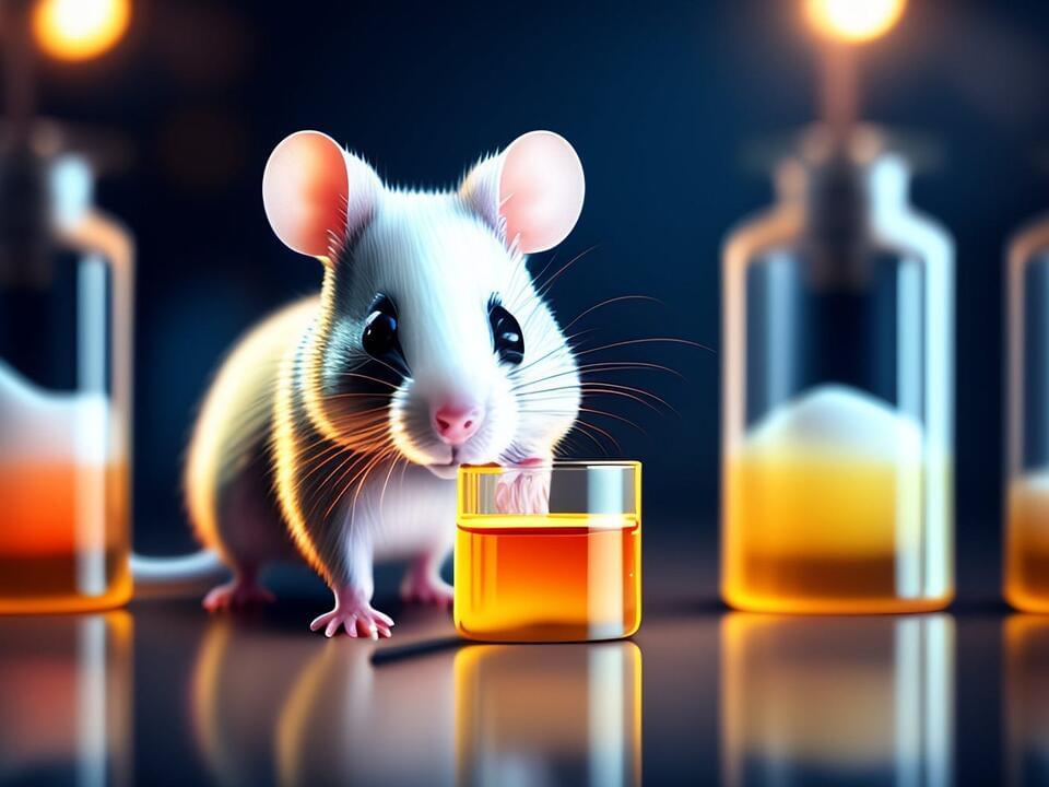
Could a fecal transplant pill be the antidepressants of the future?
Depression is real, and it is complex. Most conditions that affect our brain chemistry are going to be complex, and there are no easy, simple answers. We can’t cure depression by just exercising more, eating better, or taking a short vacation to recharge (although there is some evidence that getting more money, especially to lift you out of poverty, helps relieve depressive symptoms).
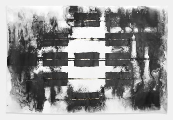
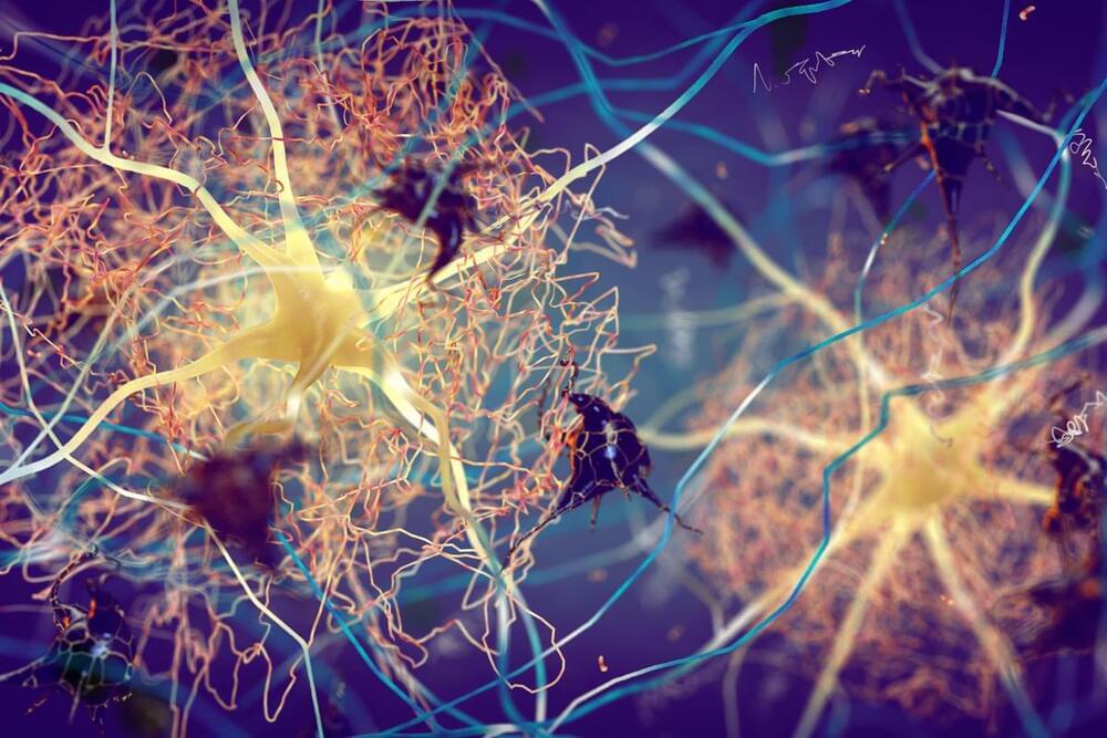
The complexity of the brain comes to life in the annual Art of Neuroscience competition.
Scattered evidence suggests that aberrant proteins act as “seeds” to transmit neurodegenerative disease, but the jury is still out.
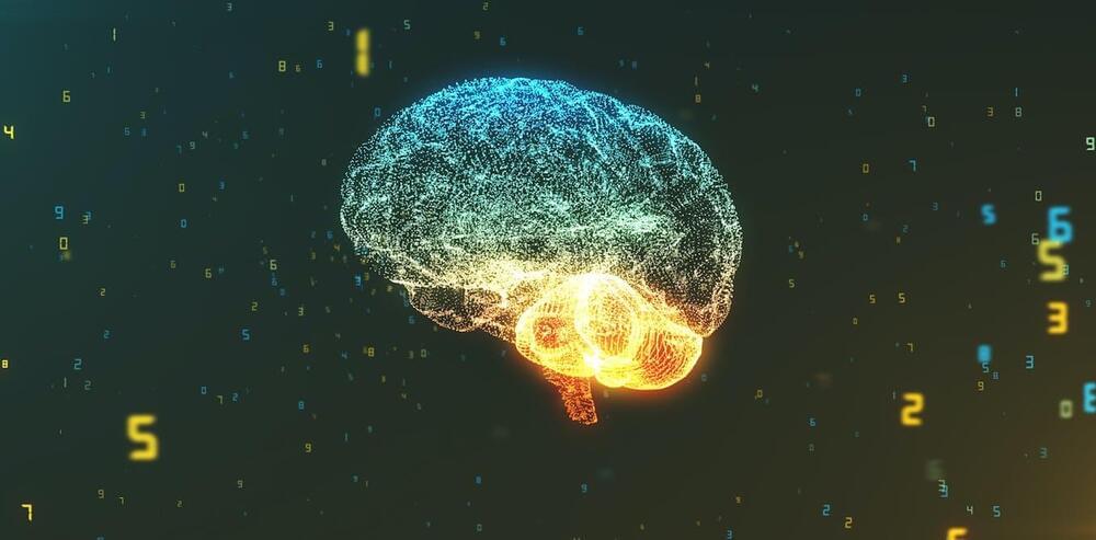
This is the concept behind mind uploading – the idea that we may one day be able to transition a person from their biological body to a synthetic hardware. The idea originated in an intellectual movement called transhumanism and has several key advocates including computer scientist Ray Kurzweil, philosopher Nick Bostrom and neuroscientist Randal Koene.
The transhumanists’ central hope is to transcend the human condition through scientific and technological progress. They believe mind uploading may allow us to live as long as we want (but not necessarily forever). It might even let us improve ourselves, such as by having simulated brains that run faster and more efficiently than biological ones. It’s a techno-optimist’s dream for the future. But does it have any substance?
The feasibility of mind uploading rests on three core assumptions.

A new venture-backed startup is capitalizing on the productivity that can be channeled while lucid dreaming, Fortune reports.
Lucid dreaming is a state of being aware that you are dreaming during your sleep cycle and the ability to control or manipulate the dream narrative. As many as 70% of people experience the phenomenon at least once in their lifetime.
Prophetic, founded earlier this year, is tapping into a new unconscious market with an innovative headpiece called the “Halo”
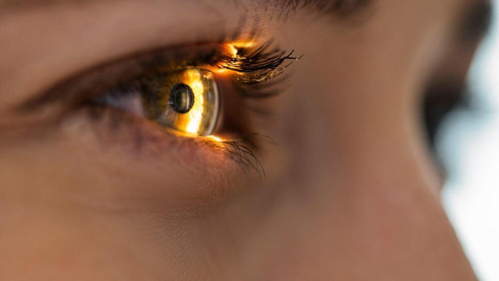
In this video, I discuss the famous “hard problem” of consciousness and why it is only a problem from the perspective of the thinking mind.
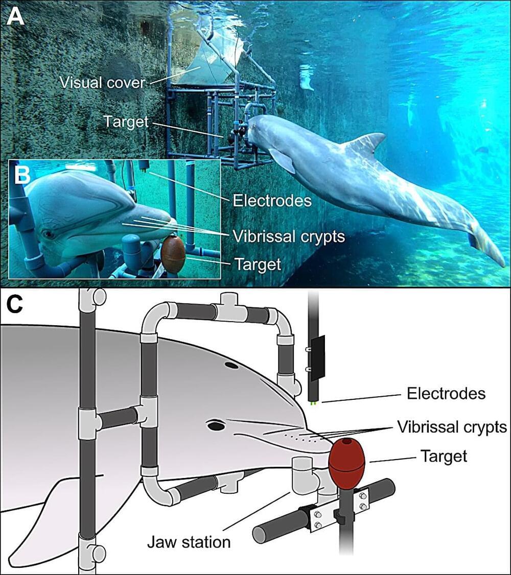
A small team of bio-scientists from the University of Rostock’s Institute for Biosciences and Nuremberg Zoo’s Behavioral Ecology and Conservation Lab, both in Germany, has found evidence that bottlenose dolphins can sense electric fields. In their study, reported in the Journal of Experimental Biology, the group tested the ability of two captive bottlenose dolphins to sense a small electric field.
Many creatures in the animal kingdom are able to sense an electric field—some sharks and the platypus, for example—but only one type of marine mammal has been found to have the ability: the Guiana dolphin. In this new effort, the research team wondered if other types of dolphins have the ability.
They chose to study bottlenose dolphins for two reasons: a pair of were available for testing at the nearby Nuremberg Zoo, and prior research suggested that neural cells in the vibrissal crypts situated along the dolphins’ snouts strongly resembled the electric-field detectors observed in sharks.
